

Med info
Diabetic retinopathy: When does it transition from monitoring to treatment?
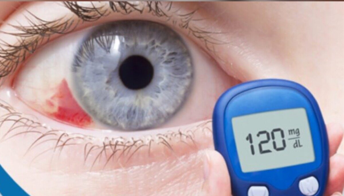
Does Every Diabetic Patient Need Retinal Treatment?
Not every person with diabetes needs active treatment for the retina, but every diabetic patient does need regular, detailed dilated fundus examinations to detect diabetic retinopathy at an early stage.
In many cases of mild to moderate diabetic retinopathy, treatment with laser or intravitreal injections is not immediately required. Instead, the ophthalmologist may opt for close follow‑up combined with strict control of blood sugar, blood pressure, cholesterol levels, and lifestyle modifications to slow the progression of the disease.
Active treatment for diabetic retinopathy becomes necessary when warning signs appear, such as retinal hemorrhages, fluid leakage involving the center of vision (diabetic macular edema), or the development of abnormal new blood vessels (neovascularization) that can seriously threaten sight.
The need for retinal treatment in a diabetic patient therefore depends on the severity of diabetic retinopathy, the findings on dilated fundus examination, optical coherence tomography (OCT), and the assessment of the retina specialist.
The key is not to wait for symptoms to appear; even if you do not need treatment right now, early detection and regular monitoring greatly reduce the risk of progression to advanced stages that require complex interventions.
The Difference Between Monitoring and Treatment — When Should You Worry?
In diabetic retinopathy, the ophthalmologist decides between simple follow‑up and active treatment based on how severely the retina is affected and how high the risk of vision loss is.
“Monitoring” means that the retinal changes are still mild or in an early stage and are not currently threatening your sight. In this case, the focus is on good control of blood sugar, blood pressure, and cholesterol, along with regular eye checks such as dilated fundus examination, retinal photography (Fundus imaging), or OCT scans every 3–6 months to watch for any progression.
Active treatment becomes necessary if there is bleeding in the retina, swelling in the macula (diabetic macular edema), proliferative diabetic retinopathy, or if there is a noticeable drop in visual acuity, sudden blurred vision, new dark floaters, or a curtain-like shadow in part of your visual field. In these situations, monitoring alone is not enough; treatment such as intravitreal injections, retinal laser therapy, or vitrectomy surgery is required, depending on the case.
You should be concerned and seek urgent assessment by an ophthalmologist or a specialized diabetic retinopathy center if you notice any sudden change in your vision between scheduled visits, or if you miss your routine dilated fundus examination. Early intervention is the single most important factor in preserving vision and reducing diabetes‑related complications in the retina.
7 Signs You’ve Moved from Monitoring to Active Treatment

Blurred or Hazy Vision
If you notice that blurred or hazy vision has become a constant rather than a temporary symptom, this is a strong indication that you’ve moved beyond the “observation only” stage and now actually need to start treatment for diabetic retinopathy.
During the observation phase, eye examinations are performed periodically while you may have little to no symptoms. However, the onset of persistent blurred vision often reflects damage to the retina caused by fluid leakage (macular edema) or mild bleeding inside the eye. This usually calls for active treatment such as intravitreal injections, retinal laser therapy, or other options that a retina specialist will determine based on the condition of your diabetic retinopathy.
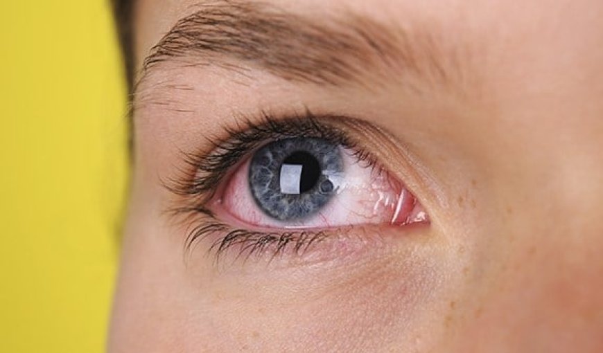
Seeing Black Spots or Floaters
Seeing black spots or what looks like flying flies (floaters) repeatedly in your field of vision may mean that your diabetic retinopathy is no longer in a stable phase that can be managed with simple monitoring.
These floaters are often caused by mild bleeding into the vitreous body inside the eye or by changes in the retinal blood vessels, and they serve as a warning sign that the disease is progressing.
Once this sign appears, your ophthalmologist’s role shifts from follow‑up and observation to formulating an actual treatment plan for diabetic retinopathy, which may involve laser photocoagulation, intravitreal injections, or even surgery (such as vitrectomy) in more advanced cases.
Difficulty Seeing at Night
When difficulty seeing at night begins to interfere with driving or moving around in dimly lit environments, this suggests that retinal damage has become more pronounced and that observation alone is no longer sufficient.
In more advanced stages, diabetic retinopathy affects the light‑sensitive cells in the retina, reducing the eye’s ability to adapt to darkness.
The clear appearance of this problem usually means you have entered a stage that requires structured treatment to slow further retinal damage and preserve the remaining vision.
Sudden Loss of Part of Your Vision
A sudden loss of vision in part of your visual field—such as not seeing one side properly or noticing a dark area in your field of view—is an urgent warning sign that usually points to advanced complications of diabetic retinopathy.
At this point, it is no longer appropriate to “wait and see” or rely on monitoring alone; treatment for diabetic retinopathy must be started immediately, as delaying therapy can result in permanent vision loss.
This type of symptom indicates that you have moved from a preventive monitoring phase into a phase that requires emergency therapeutic intervention, which may include intravitreal injections, laser treatment, or surgery depending on your doctor’s assessment.
Fluctuating Clarity of Vision
If you start noticing that your vision fluctuates during the day—sometimes you see clearly, and at other times you experience blurring or difficulty focusing—this often points to changes in retinal thickness or fluid accumulation due to diabetic retinopathy.
During the observation stage, vision is relatively stable. Persistent fluctuation, however, is a sign that the retinal blood vessels are becoming more damaged, making it necessary to move from mere follow‑up to an active treatment plan aimed at stabilizing your vision and preventing further deterioration.
Seeing Wavy or Distorted Lines
Perceiving straight lines as wavy or bent—especially when reading text or looking at door frames and window edges—may indicate fluid leakage or swelling in the macula (the central area of the retina responsible for sharp vision) as a result of diabetic macular edema.
This particular sign often means that the disease is no longer in its early, observation‑only stage, but has progressed to a phase that requires specialized treatment, such as intravitreal anti‑VEGF injections or macular laser therapy, to preserve visual acuity as much as possible.
Noticing such visual distortions is a clear indicator that you have shifted from the monitoring phase to the stage of active treatment.
Difficulty Reading or Focusing on Details
When you begin to struggle with reading books, viewing your phone screen, or focusing on small details—even while using your usual glasses—this may be a sign that diabetic retinopathy has affected your central vision.
Previously, your doctor may have relied on regular monitoring only, but this newly developed difficulty often reflects retinal changes that require actual intervention to prevent further visual decline.
At this stage, treatment for diabetic retinopathy becomes a necessity rather than an option. The primary goals are to improve your ability to read, maintain your quality of daily life, and reduce the risk of long‑term vision loss.
Stages of Diabetic Retinopathy (in simplified terms for search intent)

Mild Non‑Proliferative Diabetic Retinopathy
In the mild stage of diabetic retinopathy, the tiny blood vessels in the retina gradually start to suffer damage as a result of long‑standing high blood sugar.
At this point, small outpouchings of the vessel walls (microaneurysms) appear, along with tiny dot‑like hemorrhages and mild hard exudates. Most patients have no noticeable symptoms and do not experience obvious visual deterioration.
Early detection of mild retinopathy is crucial, because good control of blood glucose, blood pressure, and cholesterol can stabilize the condition and prevent progression to more serious stages.
This stage is therefore considered a golden window to “treat diabetic retinopathy” early, through regular follow‑up with an ophthalmologist and performing a dilated fundus examination at least once a year.
Moderate Non‑Proliferative Diabetic Retinopathy
Moderate retinopathy represents the next step in the progression of diabetic retinopathy, with more pronounced damage to the retinal microvasculature compared with the mild stage.
In this phase, retinal hemorrhages become more numerous, and cotton‑wool spots (a sign of retinal ischemia) appear. Some patients may begin to notice mild visual disturbance, especially blurred vision when reading or focusing on fine details.
The risk of progression to more severe retinopathy increases if blood glucose is not adequately controlled. At this point, the physician may start considering more targeted interventions, such as closer monitoring for diabetic macular edema or planning laser treatment when indicated.
Adhering to the diabetes treatment plan, together with regular visits to the ophthalmologist, plays a major role in slowing disease progression and preventing advancement to the more advanced stages of diabetic retinopathy.
Severe Non‑Proliferative Diabetic Retinopathy
In severe non‑proliferative diabetic retinopathy, retinal damage becomes extensive and clearly evident, with larger areas of the retinal vasculature affected and more widespread ischemic zones (poor blood supply).
At this stage, the likelihood of progression to the dangerous proliferative form increases significantly, as the ischemic retina begins to “signal for help” by stimulating the formation of new, abnormal blood vessels.
Patients may experience a marked decrease in visual acuity or persistent blurred vision, and clinically significant macular edema may develop, directly compromising central vision.
This phase usually requires an active treatment strategy within the broader management of diabetic retinopathy. Treatment may include retinal laser photocoagulation and intravitreal injections (such as anti‑VEGF agents), in addition to strict control of blood glucose and blood pressure, to reduce the risk of transition to proliferative retinopathy.
Proliferative Diabetic Retinopathy (the Most Severe Stage)
Proliferative diabetic retinopathy is the most advanced and vision‑threatening stage of diabetic retinopathy, and can lead to severe, sometimes irreversible, visual loss if not treated promptly.
During this stage, fragile, abnormal new blood vessels grow on the surface of the retina and optic disc as a response to profound retinal ischemia.
These neovascular vessels are highly prone to bleeding into the vitreous cavity, which can cause sudden onset of dark floaters, the appearance of a dark curtain across the visual field, or acute, severe loss of vision.
In addition, associated fibrovascular proliferation can exert traction on the retina, potentially resulting in tractional retinal detachment that may require surgical repair.
Proliferative retinopathy calls for intensive treatment as a core component of “diabetic retinopathy management,” including extensive panretinal laser photocoagulation, intravitreal injections, and sometimes pars plana vitrectomy to clear vitreous hemorrhage or relieve tractional membranes.
Timely diagnosis and intervention before reaching this proliferative stage remains the single most important factor in preserving vision and avoiding the serious complications of diabetic retinopathy.
Treatment Options for Diabetic Retinopathy (Aligned With Research Intent)
Intravitreal Injections
Intravitreal injections are among the most important treatment options for diabetic retinopathy, particularly when diabetic macular edema (swelling in the central part of the retina responsible for sharp vision) is present.
This approach relies on injecting anti–vascular endothelial growth factor (anti‑VEGF) agents or corticosteroids into the vitreous cavity to reduce vascular leakage, decrease macular swelling, and improve visual acuity.
The procedure is performed in the clinic under local antiseptic preparation and anesthesia, and patients usually require a series of injections at intervals determined by the ophthalmologist based on retinal response and visual improvement.
This type of treatment helps slow the progression of diabetic retinopathy and lowers the risk of vision loss; however, it does not replace the need for strict control of blood glucose, blood pressure, and cholesterol levels, which remain essential for long‑term success in managing diabetic retinopathy.
Retinal Laser Treatment
Retinal laser therapy is widely used as a primary option in the management of both non‑proliferative and proliferative diabetic retinopathy, especially when there are areas of leakage or abnormal new vessel growth.
Laser treatment works by sealing and cauterizing fragile, leaking retinal blood vessels and can also reduce the risk of vitreous hemorrhage or retinal detachment in more advanced stages, thereby preserving vision as much as possible.
Laser sessions are carried out in the clinic under local anesthesia, and multiple sessions may be required depending on the severity of diabetic retinopathy and the extent of retinal changes seen on fundus examination.
Although laser treatment usually does not restore vision that has already been lost, it is an effective tool to limit disease progression and protect the remaining vision.
Surgery (Vitrectomy)
Pars plana vitrectomy is an advanced surgical option for diabetic retinopathy, reserved for serious complications such as dense vitreous hemorrhage or retinal detachment.
During this surgery, the ophthalmic surgeon removes the blood‑filled or opacified vitreous gel, clears membranes and traction from the retinal surface, treats bleeding sites or fibrous bands, and may perform endolaser photocoagulation to stabilize the retina and reduce the risk of recurrent bleeding.
Vitrectomy is performed in the operating room under local or general anesthesia, depending on the patient’s condition. It is a delicate procedure aimed at restoring as much vision as possible or at least preventing further deterioration.
This surgical option is an important component of the treatment strategy for advanced diabetic retinopathy, but it does not eliminate the need for regular follow‑up and tight glycemic control to reduce the likelihood of future surgical interventions.
What happens if treatment is delayed?
Delaying treatment for diabetic retinopathy can cause the disease to progress significantly, shifting from the early, often symptom-free stages to advanced diabetic retinopathy. This raises the risk of serious complications such as vitreous hemorrhage, retinal detachment, or diabetic macular edema, which directly affects the center of vision.
Over time, and especially with poorly controlled blood sugar in the absence of regular eye check-ups, the tiny blood vessels in the retina can sustain irreversible damage. This may lead to blurred vision, the appearance of dark spots or floaters, difficulty reading and driving, and in advanced stages, permanent loss of sight.
Neglecting treatment or postponing a visit to a retina specialist means missing the optimal window for early intervention with therapies such as retinal laser treatment, intravitreal injections, or vitreoretinal surgery. These options are highly effective at preserving vision or significantly slowing its deterioration when started at the right time.
Early diagnosis and regular follow-up not only protect your eyes from serious complications, but also reduce the overall cost and complexity of managing diabetic retinopathy. They help you maintain your quality of life and independence for as long as possible.
Best Retina Specialist in Jeddah
Dr. Waddah Jalabi – Consultant Retina Specialist & Surgeon
Dr. Waddah Jalabi is regarded as one of the leading choices for patients seeking the best retina specialist in Jeddah for managing diabetic retinopathy across all stages.
He brings extensive clinical and surgical experience in diagnosing and treating complex retinal diseases, with a particular focus on diabetic retinopathy cases that require close monitoring and a personalized treatment plan for each patient.
Dr. Waddah utilizes state-of-the-art imaging technologies, including fluorescein angiography and optical coherence tomography (OCT), to precisely assess the status of the retinal blood vessels before formulating a tailored management strategy.
This level of precision and expertise makes consulting Dr. Waddah an excellent option for anyone in Jeddah seeking comprehensive retinal care, especially individuals with diabetes who wish to safeguard their vision and avoid serious complications.
Start Your Treatment Journey Today with Leading Retina Specialists at Batal Specialty Center
Begin managing diabetic retinopathy today at Batal Specialty Center with a distinguished team of board‑certified retina specialists. We provide precise diagnosis using state-of-the-art imaging technologies, including fluorescein fundus angiography and optical coherence tomography (OCT), to detect early changes in the retinal blood vessels.
Our ophthalmology team follows the latest international guidelines for the treatment of diabetic retinopathy, offering intravitreal injections (anti‑VEGF agents and corticosteroids), retinal laser therapy, and minimally invasive vitreoretinal surgery when indicated—aiming to halt disease progression and preserve as much visual acuity as possible.
Book your appointment now at the Retina Clinics of Batal Specialty Center to start a personalized treatment plan tailored to your overall health status and blood sugar control. Early diagnosis and regular follow‑up are key to preventing diabetic retinopathy complications and safeguarding your vision over the long term.


