

Med info
Retinal vessel occlusion
What is Retinal Vascular Occlusion?
Retinal vascular occlusion is an acute eye condition that occurs when blood flow is suddenly blocked in one of the arteries or veins supplying the retina — the light‑sensitive layer at the back of the eye responsible for image formation and clear vision.
When this blockage happens, the retinal tissue is deprived of oxygen and nutrients, leading to damage of retinal cells and symptoms such as sudden blurred vision, loss of part of the visual field, or even complete loss of vision in the affected eye.
The most common types are retinal vein occlusion (RVO) and retinal artery occlusion (RAO). These are frequently associated with risk factors such as high blood pressure, diabetes, elevated cholesterol levels, smoking, and blood clotting disorders.
Understanding retinal vascular occlusion is crucial, as it is considered an ocular emergency that requires immediate assessment by an ophthalmologist to preserve as much visual function as possible and to minimize long‑term complications.
What are the types of retinal vascular occlusion?
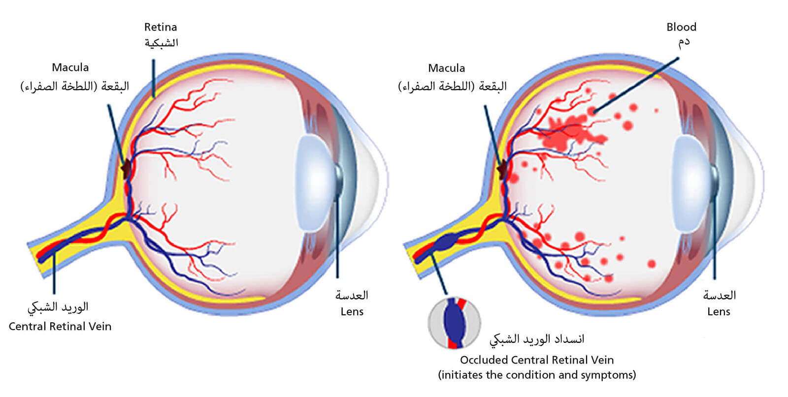
Central Retinal Vein Occlusion (CRVO)
Central retinal vein occlusion is one of the most important types of retinal vascular occlusion. It occurs when the main vein draining blood from the retina becomes blocked or thrombosed.
This blockage leads to venous congestion within the retina, fluid leakage, and retinal hemorrhages, resulting in a sudden blurring of vision that may sometimes progress to a marked loss of visual acuity.
CRVO is commonly associated with risk factors such as hypertension, diabetes mellitus, hyperlipidemia, and increasing age.
Early detection of CRVO and prompt management of complications such as macular edema are crucial for preserving as much vision as possible.
Branch Retinal Vein Occlusion (BRVO)
Branch retinal vein occlusion is another form of retinal vascular occlusion, affecting one of the smaller branches of the retinal vein rather than the main trunk.
This type of occlusion typically causes partial visual field loss or blurring in a specific area of the visual scene, depending on the location of the affected venous branch within the retina.
BRVO is frequently seen in patients with hypertension or other vascular disorders, and it may lead to fluid accumulation in the macular region (macular edema), which directly impairs central visual clarity.
Timely diagnosis of BRVO and careful follow‑up help limit complications and preserve retinal function as much as possible.
Central Retinal Artery Occlusion (CRAO)
Central retinal artery occlusion is among the most serious types of retinal vascular occlusion and is considered an ophthalmic emergency, as it usually causes sudden, profound vision loss in one eye.
CRAO results from thrombosis or obstruction of the main artery supplying the retina with oxygen and nutrients, and is often associated with atherosclerosis, cardiovascular disease, or coagulation disorders.
Severe acute ischemia leads to rapid damage of retinal cells if not treated very early; therefore, CRAO is regarded as an urgent condition requiring immediate evaluation of both the eye and the cardiovascular system.
Recognizing CRAO as a form of retinal vascular occlusion helps patients appreciate its severity and the need to seek urgent medical attention.
Branch Retinal Artery Occlusion (BRAO)
Branch retinal artery occlusion involves obstruction of one of the smaller branches of the central retinal artery. It is less common than CRAO but remains clinically significant.
BRAO typically causes sudden loss of vision in a segment of the visual field, such as a dark spot or a localized area of reduced vision, depending on the site of the affected arterial branch.
The underlying cause is often small emboli or plaques originating from larger arteries, such as the carotid arteries, or from the heart, making BRAO an important indicator of systemic vascular disease that warrants further investigation.
Early diagnosis of BRAO allows for proper assessment of stroke and cardiac risk and supports the development of a preventive treatment plan to reduce the likelihood of future vascular occlusive events.
What Are the Causes and Risk Factors of Retinal Vascular Occlusion?
Diabetes and Hypertension
Diabetes mellitus and high blood pressure are among the leading causes of retinal vascular occlusion and the most important risk factors for developing it.
When blood glucose remains elevated for prolonged periods, it damages the walls of the tiny retinal blood vessels, making them fragile and more prone to narrowing or blockage due to lipid deposits or microthrombi.
Chronic hypertension, on the other hand, leads to arterial stiffening and thickening of the vessel wall, which narrows the lumen and disrupts blood flow to the retina.
Over time, the coexistence of diabetes and hypertension significantly increases the risk of retinal vein occlusion (RVO) or retinal artery occlusion (RAO), especially when treatment is poorly controlled or regular medical follow‑up is neglected.
For that reason, people with diabetes and hypertension are strongly advised to keep their blood sugar and blood pressure within target levels and to have regular eye examinations to detect any early changes in the retinal vasculature.
Cardiovascular Disease and Circulatory Disorders
Cardiovascular diseases and circulatory disorders are key contributors to retinal vascular occlusion and are considered major associated risk factors.
Cardiac problems such as atrial fibrillation, cardiomyopathy (weak heart muscle), or valvular heart disease can promote the formation of intracardiac thrombi. These clots may then embolize through the circulation to the retinal arteries, causing sudden arterial occlusion.
Systemic atherosclerosis and impaired blood flow throughout the body likewise increase the likelihood of arterial or venous occlusion in the retina, particularly in older adults or individuals with uncontrolled hypertension or hyperlipidemia.
Therefore, controlling cardiovascular risk factors—such as smoking, obesity, physical inactivity, and elevated blood lipids—is an essential component of preventing retinal vascular occlusion and preserving vision in the long term.
Coagulation Disorders and Hypercholesterolemia
Coagulation disorders and elevated blood cholesterol play a direct role in increasing the risk of retinal vascular occlusion and its associated complications.
When the blood is more viscous or more prone to clotting—because of platelet disorders or abnormalities in coagulation factors—small thrombi form more readily and may lodge in retinal arteries or veins, causing an acute interruption of blood flow.
Raised cholesterol and triglyceride levels contribute to lipid deposition within the walls of small vessels, leading to their narrowing and sclerosis and making them more susceptible to occlusion, particularly in delicate vascular beds such as the retinal circulation.
Individuals with a family history of thrombophilia or poorly controlled hypercholesterolemia are at substantially higher risk of retinal vein occlusion or retinal artery occlusion.
Regular screening of blood lipids, adherence to a healthy diet, and appropriate medical treatment are recommended to reduce the risk of these eye and retinal complications.
Glaucoma and Other Ocular Diseases
Glaucoma (elevated intraocular pressure) and certain chronic eye diseases are associated with an increased risk of retinal vascular occlusion and are considered local factors that directly affect intraocular circulation.
In glaucoma, raised intraocular pressure exerts additional stress on the optic nerve fibers and the fine retinal vessels, compromising blood flow and making these vessels more prone to narrowing, collapse, or occlusion—particularly the retinal veins.
Other ocular conditions, such as retinal vasculitis or retinopathy secondary to systemic disease, can alter the structure of vessel walls, causing loss of elasticity and impaired autoregulation of blood flow, which in turn increases the likelihood of vascular blockage.
Regular follow‑up with an ophthalmologist for patients with glaucoma or any chronic eye disease is crucial to detect early signs of circulatory disturbance in the retina and to reduce the risk of retinal vascular occlusion and sudden vision loss.
What are the symptoms of retinal vessel occlusion?
Sudden or Gradual Vision Loss
Sudden or gradually progressive loss of vision is one of the key signs of retinal vascular occlusion, and it may affect one eye or both, depending on the type of blockage.
In central or branch retinal artery occlusion (CRAO/BRAO), vision loss is typically abrupt and severe, developing over minutes to hours. Patients often describe it as if a “black curtain” has fallen over the eye.
In retinal vein occlusion (central or branch RVO), visual decline is more likely to be gradual, over hours or days, and the patient may notice increasing difficulty with reading or seeing fine details.
In both situations, any sudden or unexplained vision loss is an emergency that requires immediate evaluation by an ophthalmologist, as delayed diagnosis and treatment of retinal vascular occlusion can result in permanent loss of sight.
Blurred Vision and Reduced Visual Acuity
Blurred vision and reduced visual acuity are common early manifestations of retinal vascular occlusion, particularly in retinal vein occlusion or occlusion of smaller retinal vessels and branches.
The patient may feel that their vision is no longer as sharp as before, colors look washed out, or straight lines appear blurred or slightly distorted.
This blurring may worsen over time or fluctuate, and it is often more noticeable when trying to focus on near tasks, such as reading or using a smartphone.
If blurred vision or reduced visual clarity persists without an obvious cause—such as eye strain or needing a change in eyeglass prescription—an eye specialist should be consulted to rule out retinal vascular occlusion or other ocular conditions.
Floaters or Dark Spots in the Field of Vision
The appearance of floaters or dark spots in the visual field can accompany some cases of retinal vascular occlusion, especially when the blockage leads to intraocular hemorrhage or fluid leakage (edema).
Patients often describe these floaters as small black dots, strands, or “flies” that drift with eye movement, becoming more noticeable against bright backgrounds such as the sky or a computer screen.
Although floaters are frequently benign, their sudden onset, a marked increase in their number, or their appearance together with flashes of light (photopsia) or blurred vision warrants urgent assessment. These signs may indicate a retinal vascular occlusion or other serious problems such as retinal tear or retinal detachment.
Loss of Part of the Visual Field
Another possible symptom of retinal vascular occlusion is loss of a portion of the visual field. The patient may perceive this as a “dark area” or a “blank spot” affecting one side of what they see.
The affected area may be in the upper, lower, or lateral part of the visual field, depending on the exact location of the occlusion within the retinal vasculature.
Sometimes the patient does not immediately notice these dark patches and only becomes aware of them when covering one eye, or while driving or reading signs, where part of the scene appears missing.
Any localized visual field defect—especially if it appears suddenly or is accompanied by blurring or pain around the eye—requires prompt evaluation by an ophthalmologist to determine whether it is due to retinal vascular occlusion, optic nerve pathology, or other retinal disease.
When Is the Condition Considered an Emergency?
Retinal vascular occlusion is considered an emergency when there is a sudden loss of vision in one or both eyes, especially if the loss develops acutely over minutes to hours, or if it is accompanied by severe visual blurring or a fixed dark spot in the visual field that does not go away.
The situation becomes more critical if the retinal vessel occlusion occurs along with other symptoms such as sudden, severe headache, difficulty speaking, or weakness and numbness in one limb, as these may indicate a high risk of stroke.
In such cases, you must go immediately to an eye emergency department or the nearest hospital without delay, because rapid medical intervention within the first few hours of a central retinal artery or vein occlusion can improve the chances of preserving remaining vision and reduce complications such as macular edema or permanent vision loss.
How Is Retinal Vascular Occlusion Diagnosed?
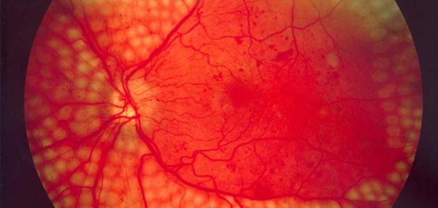
Diagnosis of retinal vascular occlusion begins with a detailed medical history, focusing on symptoms such as blurred vision or sudden vision loss in one eye. The ophthalmologist then performs a dilated fundus examination using a specialized ophthalmoscope to obtain a clear, magnified view of the retina and its blood vessels.
Fluorescein angiography is frequently ordered to visualize the retinal circulation, pinpoint the site of the occlusion, and assess how severely blood flow in the retinal arteries or veins has been compromised.
Optical coherence tomography (OCT) is also used to evaluate retinal thickness and detect intraretinal fluid or macular edema, particularly in the macula, which is responsible for central visual acuity.
In some cases, intraocular pressure is measured and other eye conditions that can mimic retinal vascular occlusion are ruled out. Laboratory tests are often requested—including blood glucose, lipid profile, coagulation studies, and blood pressure assessment—to identify systemic risk factors that may have contributed to the occlusion and to develop a comprehensive treatment plan aimed at preventing recurrence.
What are the treatment options for retinal vascular occlusion?
Intraocular Anti‑VEGF Injections
Intravitreous injections of anti‑vascular endothelial growth factor (Anti‑VEGF) agents are among the main treatment options for retinal vein occlusion, especially when there is macular edema and fluid accumulation at the center of vision.
These medications reduce the leakage from abnormal blood vessels and decrease retinal swelling, which helps improve visual acuity or at least prevent further deterioration.
Anti‑VEGF drugs are injected into the vitreous cavity under simple local anesthesia in the clinic, and patients usually require a series of injections at regular intervals, depending on how the retina responds to treatment.
The ophthalmologist decides on the need for this type of therapy in retinal vein occlusion after assessing macular thickness using optical coherence tomography (OCT) and performing a dilated fundus examination.
Retinal Laser Therapy
Retinal laser treatment is an important modality in the management of retinal vein occlusion, particularly in cases with extensive areas of ischemia or when abnormal neovascularization is present.
Laser photocoagulation targets and seals damaged retinal areas or fragile blood vessels, thereby reducing the risk of intraocular hemorrhage and serious complications such as neovascular glaucoma.
Laser may be used alone or in combination with intravitreal Anti‑VEGF injections or corticosteroids. The ophthalmologist selects the type of laser and designs the treatment plan according to the type and location of the vein occlusion and the degree of visual involvement.
Intraocular Steroid Injections
Intraocular corticosteroid therapy (such as intravitreal triamcinolone injections or long‑acting steroid implants) is an additional treatment option in retinal vein occlusion when associated with chronic macular edema or edema that is resistant to other therapies.
Steroids act by reducing inflammation and retinal swelling, which can improve or stabilize vision; however, they may be associated with side effects such as elevated intraocular pressure or cataract formation.
For this reason, steroid treatment is chosen carefully and is often reserved for specific patient groups or when retinal vein occlusion does not show adequate response to Anti‑VEGF therapy alone.
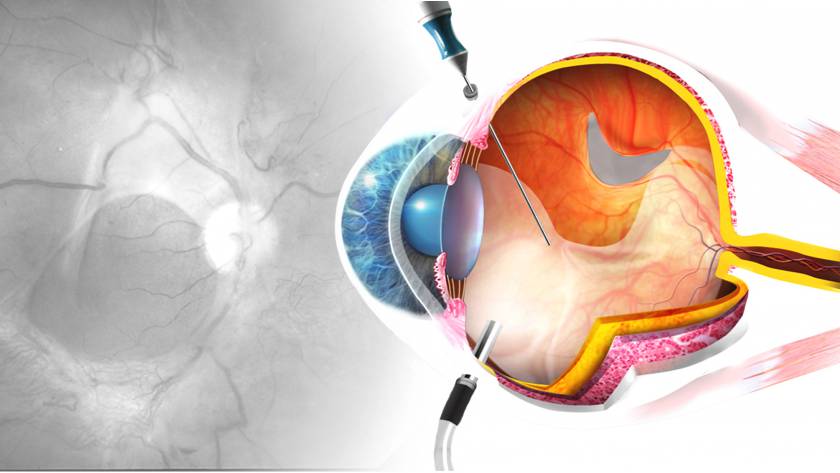
When Is Surgery Necessary?
Surgery becomes a consideration for retinal vein occlusion in selected situations, particularly when advanced complications develop, such as dense, non‑resolving vitreous hemorrhage or tractional retinal detachment caused by abnormal new vessels and fibrous membranes.
In such cases, the vitreoretinal surgeon may perform pars plana vitrectomy to clear the hemorrhage, remove tractional membranes, reattach the retina, and sometimes combine this with endolaser photocoagulation.
Most patients with retinal vein occlusion do not require surgery; however, it becomes necessary when these complications threaten retinal integrity and visual stability and fail to respond to medical treatment or laser therapy.
Can vision be restored after a retinal vascular occlusion?
The potential to regain vision after a retinal vascular occlusion depends on several factors: the type of occlusion (arterial vs. venous), the extent of retinal damage, and how quickly treatment is initiated.
In some cases of branch or partial retinal vein occlusion, visual acuity may gradually improve with appropriate treatment, such as intravitreal anti-VEGF injections or corticosteroids, which help reduce macular edema and improve retinal perfusion.
Central retinal artery occlusion, on the other hand, is usually more serious. It often causes a sudden, profound loss of vision, and the chances of visual recovery are limited if medical intervention is not provided within the first few hours of onset.
Therefore, seeking urgent ophthalmic evaluation at the first sign of sudden blurred vision or loss of part of the visual field is critical to preserving remaining sight and minimizing the long-term complications of retinal vascular occlusions.
What are the possible complications if the condition is left untreated?
Neglecting the treatment of retinal vascular occlusion can lead to serious, permanent damage to the eye and vision.
A blockage in the retinal vein or artery can cause irreversible injury to retinal cells, resulting in severe visual impairment or sudden, permanent loss of vision in the affected eye.
If retinal vessel occlusion persists without proper intervention, several complications may develop, such as macular edema (swelling of the macula responsible for sharp central vision), and the growth of fragile, abnormal new blood vessels inside the eye. This significantly increases the risk of vitreous hemorrhage and elevated intraocular pressure due to neovascular glaucoma—conditions that may ultimately result in complete blindness.
Patients may also experience persistent blurred vision, along with difficulty reading, driving, and performing daily tasks, which can greatly reduce their quality of life.
For these reasons, early diagnosis and regular follow‑up with a retina specialist or ophthalmologist are essential to limit the complications of retinal vascular occlusion and preserve as much visual function as possible.
How can retinal vascular occlusion be prevented?
Preventing retinal vascular occlusion starts with tight control of the risk factors that damage the small blood vessels in the eye, such as hypertension, diabetes, and high cholesterol.
Good control of blood glucose, blood pressure, and blood lipids is essential to protect the retinal vasculature from atherosclerosis and thrombosis. Regular follow‑up with an internist or cardiologist and adhering to prescribed medications are strongly recommended.
Quitting smoking, reducing salt and saturated fat intake, and exercising regularly all help improve circulation and lower the likelihood of blockages in the retinal arteries and veins.
Regular eye examinations with an ophthalmologist—especially for people with diabetes, high blood pressure, cardiovascular disease, or a family history of retinal vascular occlusion—are crucial to detect any early retinal changes before they progress to a complete occlusion.
Maintaining a healthy body weight, staying well hydrated, and treating coagulation disorders or atrial fibrillation under medical supervision are additional preventive measures that reduce the risk of retinal vascular occlusion and help preserve vision as much as possible.
Best Retina Specialist in Jeddah
.
Dr. Waddah Jalabi and His Expertise in Retinal Disease Management
Dr. Waddah Jalabi is widely recognized as one of the leading retina specialists in Jeddah, particularly for patients seeking expert care for retinal vascular occlusions, where precise experience and early diagnosis are critical.
He has extensive experience in managing complex retinal disorders, including branch and central retinal vein occlusion (RVO) as well as retinal artery occlusion (RAO), and follows up‑to‑date, evidence‑based treatment protocols designed to preserve visual acuity as much as possible.
Dr. Jalabi utilizes state-of-the-art diagnostic technologies such as optical coherence tomography (OCT) and fluorescein angiography to accurately assess the extent of retinal damage and tailor an individualized treatment plan for each patient.
This high level of subspecialty expertise, combined with his focus on long-term follow‑up and monitoring, has made him a preferred choice for patients seeking advanced retinal care in Jeddah, especially those with diabetic retinopathy or hypertension-related complications that can lead to retinal vascular occlusions.
Book an urgent assessment at Batal Eye Specialist Center
If you experience sudden blurred vision or partial/complete loss of sight, this may indicate retinal vascular occlusion—an ocular emergency that requires immediate evaluation by a retina specialist.
At Batal Eye Specialist Center, our board‑certified consultants have extensive expertise in diagnosing and managing retinal vein occlusion (RVO) and retinal artery occlusion (RAO) using state‑of‑the‑art imaging modalities, including Optical Coherence Tomography (OCT) and Fluorescein Angiography (FA) for detailed retinal vascular assessment and accurate diagnosis.
Schedule an urgent assessment now to promptly determine the cause of your visual deterioration and initiate an individualized treatment plan aimed at reducing the risk of complications such as permanent vision loss. We also provide structured follow‑up for patients with diabetes, hypertension, and hyperlipidemia, given their higher risk of retinal vascular occlusion.
You can book your appointment easily via direct phone call to the center or through online booking. Your case will be prioritized and triaged as an emergency in the retina clinic.


