

Med info
Corneal examination before LASIK: What does it reveal?
Why Is Corneal Assessment Before LASIK Essential?
A thorough corneal assessment before LASIK is a critical step to ensure both the safety and success of the procedure. It goes far beyond simply measuring the degree of refractive error; it also evaluates corneal thickness, curvature, and surface regularity to confirm that the cornea can safely tolerate the removal of a thin layer of tissue during vision correction.
Pre‑LASIK corneal evaluation allows early detection of subtle problems such as keratoconus or subclinical corneal thinning, conditions that can lead to serious complications like corneal ectasia or distorted vision if not identified beforehand.
This assessment also helps the surgeon choose the most appropriate technique for each patient, determine the safe amount of correction, or even rule out LASIK entirely and recommend safer alternatives such as surface ablation procedures (like PRK) or phakic intraocular lenses.
Undergoing a meticulous corneal examination in a reputable eye center is therefore the cornerstone for achieving stable vision, long‑lasting results, and the lowest possible risk of complications.
Essential Corneal Tests Before LASIK
Corneal Topography and What It Reveals
Corneal topography is one of the most important pre‑LASIK tests. It creates a detailed map of the curvature of the anterior corneal surface, accurately showing flatter and steeper areas.
This examination helps the ophthalmologist detect any irregularities in corneal shape, such as keratoconus or its early stages, which may render LASIK unsafe or make another form of vision correction more appropriate.
Topography also assesses how regular the corneal surface is, which directly affects the visual accuracy after LASIK and helps in selecting the most suitable excimer laser ablation profile.
With this information, the risk of complications can be reduced and the chances of achieving stable, clear vision after surgery are improved.
Corneal Pachymetry and Its Importance
Corneal pachymetry is an essential pre‑LASIK test that accurately measures corneal thickness in microns.
The safety of LASIK depends on having sufficient corneal tissue to allow laser ablation while maintaining an adequate residual stromal bed, in order to prevent postoperative corneal weakening or ectasia.
If the cornea is thinner than the recommended limit, the doctor may advise alternative procedures such as surface ablation (PRK) instead of conventional LASIK, or may decide against laser vision correction altogether.
Pachymetry therefore helps tailor the treatment plan to each patient and ensures the highest possible level of safety and long‑term visual stability.
Anterior and Posterior Corneal Evaluation (Pentacam / Orbscan)
Assessment of the cornea with devices such as Pentacam or Orbscan is among the most precise pre‑LASIK investigations, as it evaluates both the anterior and posterior corneal surfaces as well as corneal thickness.
This three‑dimensional imaging detects subtle changes on the posterior corneal surface that are not visible with conventional tests, helping to identify early keratoconus or biomechanical instability.
It also provides data on the distribution of corneal thickness, anterior chamber depth, and higher‑order aberrations—information that is critical for successful LASIK planning.
Using this technology, the surgeon can more accurately judge the suitability of the cornea for LASIK and reduce the likelihood of complications such as postoperative corneal ectasia.
Corneal and Anterior Segment OCT

Anterior segment optical coherence tomography (OCT) is a high‑resolution imaging modality that uses light waves to obtain cross‑sectional images of the cornea and surrounding structures.
As part of the basic pre‑LASIK workup, OCT helps measure the thickness of individual corneal layers, evaluate the integrity of the epithelium and stroma, and assess anterior chamber depth and angle configuration.
This information is particularly valuable in thin corneas, in eyes with previous surgery, or where ocular surface disease is suspected, as it enables safer decisions regarding the type and depth of laser vision correction.
OCT is also useful in postoperative follow‑up to monitor the corneal flap, ensure tissue clarity, and confirm long‑term corneal stability after LASIK.
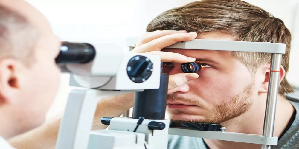
Slit‑Lamp Examination
Slit‑lamp biomicroscopy is a fundamental clinical examination performed by the ophthalmologist to assess the cornea and anterior segment directly under magnification and focused illumination.
Through this exam, the doctor can identify superficial or deep corneal opacities and scars, and detect conjunctival or eyelid inflammation or allergy that may compromise corneal healing after LASIK.
The slit‑lamp exam also allows evaluation of tear film quality, the crystalline lens, and other anterior segment structures to rule out conditions that could affect vision.
This step is indispensable in the pre‑LASIK corneal assessment, as it correlates instrument‑based findings with direct clinical examination, leading to a more accurate diagnosis and a safer treatment plan.
Dry Eye Assessment (Schirmer Test)
The Schirmer test is a simple yet important component of the basic pre‑LASIK evaluation, used to quantify tear production over a set period of time.
It helps diagnose dry eye before surgery—a factor that can significantly influence patient comfort and visual quality after LASIK, since the laser procedure itself can temporarily worsen dryness.
If the Schirmer test shows reduced tear secretion, the ophthalmologist may recommend treating dry eye first, using lubricating eye drops or other therapies, before proceeding with LASIK.
Addressing dry eye in advance helps reduce postoperative symptoms such as burning, stinging, and fluctuating vision, thereby improving both the patient experience and the overall outcome of laser vision correction.
Inclusion and Exclusion Criteria for LASIK in Line with the Study Objectives
Minimum acceptable corneal thickness
Corneal thickness is one of the most important criteria for eligibility for LASIK, since the excimer laser works by removing part of the corneal tissue to reshape it and correct refractive errors.
In general, a minimum preoperative corneal thickness is required (commonly around 500 microns), while ensuring that a residual stromal bed of at least 250–300 microns is preserved after LASIK. This safety margin helps reduce the risk of postoperative corneal weakening or progressive thinning.
If the cornea is thinner than the safe range, the surgeon may advise against LASIK and recommend alternatives such as surface ablation procedures (like PRK) or phakic intraocular lenses, in order to protect long‑term ocular health.
Stable refraction before surgery
One of the key prerequisites for LASIK candidacy is confirming that the refraction (degree of myopia, hyperopia, and astigmatism) has been stable for at least about one year.
Ongoing changes in refractive error indicate that the eye is still undergoing shifts, which means that visual defects may recur after LASIK or that an enhancement procedure might be needed.
During the pre‑LASIK assessment, the ophthalmologist reviews previous refractions and compares them with the current measurements. If there is a significant discrepancy, surgery is postponed until the prescription stabilizes, to achieve more accurate and long‑lasting results.
Normal vs. abnormal corneal curvature
Corneal curvature, usually assessed by corneal topography or tomography, is a critical factor in deciding whether a patient is suitable for LASIK.
In a normal cornea, the curvature is regular and smooth, allowing the laser to reshape the surface accurately and achieve stable postoperative vision.
In contrast, in cases of abnormal curvature—such as early keratoconus or clear irregularities on the topography map—the risk of complications increases, including progression of astigmatism or deterioration of visual acuity over time.
Therefore, any signs of abnormal corneal curvature may render a patient unsuitable for LASIK, and safer alternative treatment options are considered based on the specific condition of each eye.
Presence of corneal disease or abnormalities
Pre‑existing corneal disease or structural abnormalities are among the most important exclusion criteria when assessing a patient’s suitability for LASIK.
These include conditions such as keratoconus, significant corneal scarring, chronic keratitis, advanced corneal thinning, or severe ocular surface disorders.
Such diseases can interfere with corneal healing after LASIK and increase the likelihood of visual deterioration, haze, or disturbing optical aberrations.
During the preoperative work‑up, the surgeon uses advanced diagnostic devices to evaluate corneal integrity and detect even subtle abnormalities. If an unstable or significant corneal pathology is identified, LASIK is ruled out and alternative, safer management strategies are considered to preserve visual quality and ocular health.
How do you prepare for corneal evaluation before LASIK?
Before having a corneal assessment for LASIK, your ophthalmologist will usually ask you to stop wearing contact lenses for a specific period—this may range from a few days for soft lenses to several weeks for rigid gas permeable (hard) lenses, as contacts can alter the corneal shape and affect the accuracy of the measurements.
On the day of the exam, it’s best to come in without any eye makeup, including eyeliner or mascara, to reduce the risk of contaminating the eye or the diagnostic instruments.
The doctor or clinical team will take a detailed medical and ocular history, including any previous eye surgeries, chronic systemic conditions such as diabetes or hypertension, and any medications you take regularly, since these factors can influence your suitability for LASIK.
You’ll also be advised to get adequate rest beforehand and to avoid prolonged eye strain from screens, so that measurements of corneal thickness, refractive error, and intraocular pressure are as precise as possible.
In some cases, eye drops may be used to dilate the pupils or lubricate the ocular surface. Because this can temporarily affect your vision, it’s wise to bring someone with you so you don’t have to drive yourself home if your sight is blurred after the examination.
By following these simple steps, you help ensure that the pre‑LASIK corneal evaluation is comprehensive and reliable, enabling your ophthalmologist to obtain the most accurate data and choose the safest, most appropriate treatment plan for your eyes.
What happens after you get your test results?
Once your pre-LASIK corneal assessment is complete, the ophthalmologist reviews all detailed measurements—such as corneal thickness, shape, surface regularity, and pupil size—to determine whether LASIK can be performed safely in your case.
If the corneal scan shows that your cornea is adequately thick, with no signs of keratoconus or biomechanical weakness, you are most likely considered a suitable candidate for LASIK. At this point, the doctor will explain your proposed treatment plan, the steps of the procedure, possible risks, and the expected degree of visual improvement.
If, on the other hand, the results show issues such as corneal thinning or irregularity, the doctor may recommend alternative vision correction options, such as SMILE (Small Incision Lenticule Extraction), implantable collamer lenses (ICL), or simply continuing with glasses or contact lenses to protect the health of your cornea.
In all cases, analyzing your corneal evaluation before LASIK helps you make an informed decision about surgery. You will either receive clear preoperative instructions if you are a good candidate, or a tailored follow‑up and eye‑protection plan if surgery is not the best option for you.
Risks of Skipping Corneal Assessment Before LASIK
Skipping a thorough corneal evaluation before LASIK can expose the patient to serious complications that could have been easily prevented with proper preoperative assessment.
The purpose of examining the cornea before LASIK is to confirm its thickness, shape, and regularity of the surface, and to detect any early signs of keratoconus or abnormal thinning that would make the eye unsuitable for laser vision correction.
When this critical step is overlooked, the risk of postoperative corneal weakening (such as keratoconus or corneal ectasia) increases, leading to progressive visual deterioration, distorted vision, and difficulty achieving good vision even with glasses or contact lenses.
The patient may also experience severe dry eye, night-time glare and halos, visual quality below expectations, and in some cases may require additional corrective procedures or even corneal transplantation in advanced stages.
For these reasons, pre‑LASIK corneal assessment—including corneal topography, pachymetry (corneal thickness measurement), and evaluation of the ocular surface—is an essential prerequisite for a safe procedure and for achieving stable, long‑term visual outcomes.
Best Specialist for Pre-LASIK Corneal Assessment in Jeddah – Dr. Issa BaEisa
Dr. Issa BaEisa is considered one of the leading ophthalmologists in Jeddah for pre‑LASIK corneal assessment. He combines extensive experience in ophthalmology and eye surgery with the use of state-of-the-art corneal topography and pachymetry devices to accurately measure corneal thickness and detect any early signs of corneal weakness or keratoconus.
Dr. BaEisa is meticulous about performing a comprehensive eye evaluation before LASIK. This includes visual acuity testing, refraction to determine the exact prescription, intraocular pressure measurement, and detailed analysis of the corneal surface and shape to ensure the long-term safety of the procedure.
With this thorough approach, he can decide whether the patient is a suitable candidate for LASIK or would be better served with alternatives such as surface ablation (PRK) or phakic intraocular lens implantation, significantly reducing the risk of post-operative complications.
So, if you are looking for the best doctor for corneal examination before LASIK in Jeddah, visiting Dr. Issa BaEisa’s clinic will provide you with a reliable assessment and a personalized treatment plan that prioritizes your eye safety and optimizes your visual outcome.
Book Your Pre-LASIK Corneal Assessment at Batal Eye Specialty Center
At Batal Eye Specialty Center, you can easily book a comprehensive pre-LASIK corneal assessment to determine how suitable your eyes are for vision correction procedures, with the highest standards of safety.
Your cornea will be thoroughly evaluated using the latest corneal tomography and topography technologies to measure corneal thickness, shape, and curvature, and to detect any conditions that might contraindicate LASIK, such as keratoconus or abnormal corneal surface changes.
This pre-LASIK corneal assessment helps you choose the most appropriate technique for your case, accurately anticipate expected outcomes, and minimize potential risks—making your vision correction journey safer and more effective.
You can now schedule your pre-LASIK corneal assessment at Batal Eye Specialty Center by phone or through online booking, and receive a precise evaluation from consultant ophthalmologists specialized in LASIK and surface laser procedures (PRK and related techniques).
By taking this step, you ensure that your decision to undergo LASIK is based on reliable medical information and a comprehensive corneal evaluation tailored to the specific characteristics and needs of each eye.


