

Med info
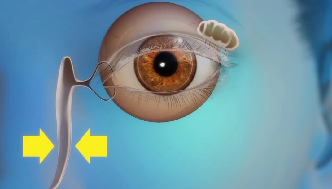
The processes of the lacrimal system: When is the cause of tears a blocked duct?
Who Needs Lacrimal System Surgery?

Lacrimal system surgery is indicated for people with blocked tear ducts or impaired tear drainage that leads to constant tearing, eye redness, recurrent infections around the eye or lacrimal sac, or mucous and purulent discharge from the inner corner of the eye.
Those who most commonly need these procedures include adults with acquired nasolacrimal duct obstruction caused by chronic inflammation, facial or nasal trauma, or previous eyelid or nasal surgery, as well as children born with congenital nasolacrimal duct obstruction who have not responded to conservative measures such as massage or medical treatment.
Lacrimal surgery may also be recommended for patients whose excessive tearing causes blurred vision or persistent discomfort that interferes with daily activities, or for those with an abscess or an acutely inflamed lacrimal sac (dacryocystitis) that requires surgical drainage and measures to prevent recurrent infection.
In all cases, the need for surgery and the most appropriate type of procedure are determined by an ophthalmologist specialized in lacrimal drainage disorders, after a thorough examination and appropriate diagnostic tests such as lacrimal irrigation (tear duct probing and syringing) or imaging studies of the nasolacrimal system.
Symptoms and Signs of Nasolacrimal Duct Obstruction
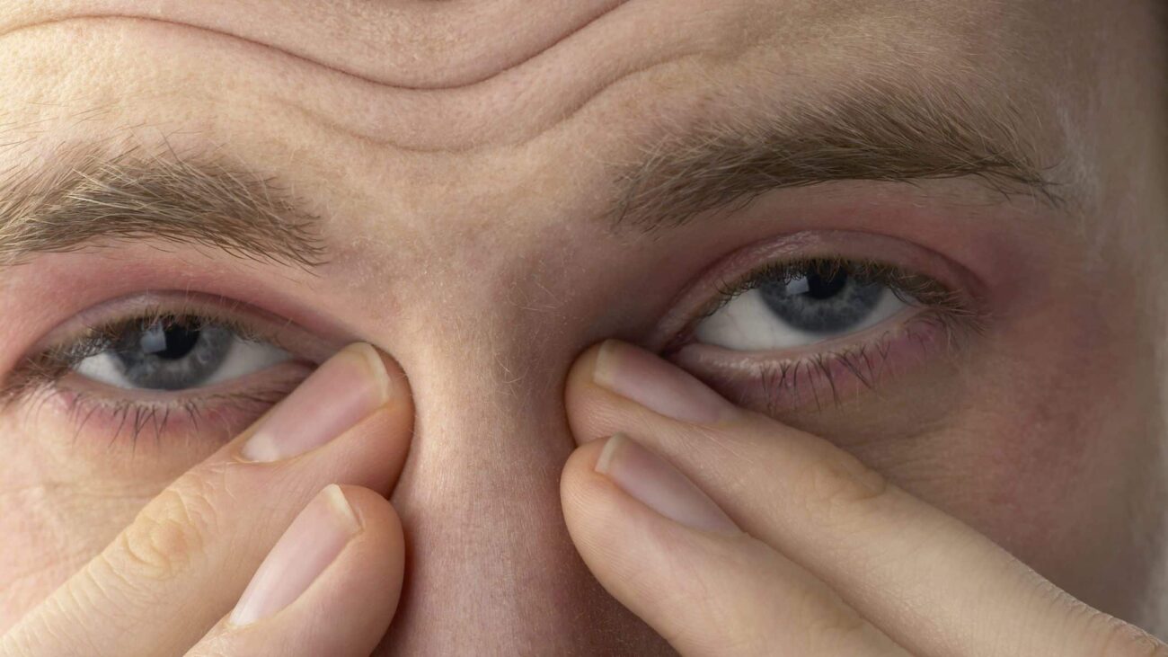
Symptoms and signs of nasolacrimal duct obstruction usually develop gradually. The most prominent is excessive tearing, known as epiphora, where tears overflow onto the cheek without an obvious cause. Patients often notice this more in cold weather or when exposed to wind.
The affected person may also experience burning, stinging, or a sensation of a foreign body in the eye, in addition to recurrent conjunctivitis or dacryocystitis (inflammation of the lacrimal sac). Dacryocystitis typically presents with redness, swelling, and pain at the inner corner of the eye near the nose, and may be accompanied by mucous or purulent discharge, especially when gentle pressure is applied over the lacrimal sac area.
In chronic cases of nasolacrimal duct obstruction, the patient may notice mild blurring of vision due to pooling of tears over the corneal surface, along with increased light sensitivity and persistent ocular irritation.
If there is sudden severe pain, marked swelling, and fever, this may indicate acute dacryocystitis, a condition that requires urgent evaluation by an ophthalmologist to establish an accurate diagnosis and determine the appropriate management plan—whether medical treatment or lacrimal drainage surgery—depending on the severity and level of obstruction.
How is nasolacrimal duct obstruction diagnosed?
Diagnosis of nasolacrimal duct obstruction begins with a detailed medical history. The ophthalmologist will ask about how long the tearing has been present, whether it is getting worse, the presence of discharge or recurrent infections, and any previous surgeries around the eyes or nose.
The doctor then examines the eyes, eyelids, and lacrimal puncta using a slit lamp to look for swelling, redness, or narrowing of the lacrimal openings.
This is followed by “tear drainage tests,” such as the fluorescein dye disappearance test. A colored dye is instilled into the eye, and its clearance through the nasolacrimal system is monitored to help determine the site of obstruction.
The physician may also use a fine cannula to irrigate the nasolacrimal duct with saline (lacrimal irrigation). Failure of the fluid to pass into the nose, or its regurgitation from the opposite punctum, indicates a blockage within the lacrimal drainage pathway.
In some cases, particularly before lacrimal surgery, imaging studies are requested, such as contrast dacryocystography, CT scans, or MRI. These help pinpoint the exact location and cause of the obstruction and guide the choice of the most appropriate treatment plan.
Types of Lacrimal System Surgeries (for research purposes)
Probing of the Nasolacrimal Duct in Children and Adults

Probing of the nasolacrimal duct is one of the most commonly performed lacrimal procedures for treating nasolacrimal duct obstruction, particularly in children.
The procedure is usually done under general anesthesia in children and under local or general anesthesia in adults. An ophthalmologist passes a fine probe through the lacrimal drainage system to open the blocked duct and restore normal tear flow.
Probing is recommended when tearing (epiphora) and recurrent eye infections persist despite conservative measures such as lacrimal sac massage and eye drops.
It is generally considered the first-line interventional step before moving on to more extensive surgery, and it helps prevent long‑term complications of nasolacrimal duct obstruction.
External Dacryocystorhinostomy (External DCR)
External dacryocystorhinostomy (External DCR) is a more advanced lacrimal surgery, typically performed when simpler methods fail to relieve nasolacrimal duct obstruction.
In this procedure, a small skin incision is made beside the nose to access the lacrimal sac and create a new passage between the lacrimal sac and the nasal cavity, allowing tears to drain more effectively.
External DCR is associated with a high success rate and is often used for chronic obstruction accompanied by recurrent infection (dacryocystitis) or lacrimal sac mucocele.
Although it is a conventional open surgery, the skin incision is small and usually heals well with proper care, making it an effective treatment option for many patients with nasolacrimal duct obstruction.
Endoscopic Dacryocystorhinostomy (Endoscopic DCR)
Endoscopic dacryocystorhinostomy (Endoscopic DCR) is a less invasive alternative to external DCR.
Using a nasal endoscope introduced through the nose, the surgeon creates a new drainage pathway between the lacrimal sac and the nasal cavity without any external skin incision.
This approach is often preferred for patients who are particularly concerned about cosmetic appearance or who have skin problems around the eye, as it avoids visible scars and may shorten recovery time.
The choice between external and endoscopic DCR depends on a joint assessment by the ophthalmologist and ENT specialist, taking into account the site and extent of the obstruction, nasal anatomy, and other individual factors.
Silicone Tube Intubation for Tear Drainage
Silicone tube intubation is an important adjunctive procedure within the spectrum of lacrimal surgeries.
A fine silicone tube (silicone stent) is threaded through the lacrimal drainage system, typically from the puncta in the eyelids into the nasal cavity, to keep the passage open during healing after probing or DCR surgery.
The tube helps prevent re‑obstruction and maintains tear drainage until the tissues around the new passage have healed and stabilized.
It is usually left in place for several weeks to a few months, then removed easily in the clinic, and can be used in both children and adults depending on the type of lacrimal procedure and the clinical condition.
Balloon Dacryoplasty (Balloon Dilatation of the Nasolacrimal Duct)
Balloon dacryoplasty is a minimally invasive technique used particularly in cases of partial obstruction or narrowing of the nasolacrimal duct.
A fine catheter is introduced into the nasolacrimal duct, carrying a small balloon that is gradually inflated to dilate the narrowed segment and re‑establish adequate tear flow.
This method can be applied in selected cases of nasolacrimal duct obstruction in both children and adults, either as an alternative to open surgery or as an adjunct following probing.
The suitability of balloon dacryoplasty is determined by the ophthalmologist based on clinical examination and lacrimal system imaging, taking into account the patient’s age and the pattern of obstruction.
Modern, Minimally Invasive Techniques for Nasolacrimal Duct Obstruction
Lacrimal surgery has evolved to include newer, less invasive techniques aimed at treating nasolacrimal duct obstruction with less pain and shorter recovery times.
These include laser-assisted endoscopic dacryocystorhinostomy (Laser DCR), ultra‑fine catheter systems, and advanced lacrimal stents designed to keep the tear drainage pathway patent for an optimal period.
Such approaches are often used in early or less severe obstruction, or in patients who are poor candidates for traditional open surgery, while maintaining effective tear drainage and reducing complication rates.
Selecting the most appropriate technique requires comprehensive evaluation by a lacrimal surgery specialist (oculoplastic / ophthalmic surgeon), to achieve the best possible outcome with the least necessary intervention.
Surgical and non-surgical management of nasolacrimal duct obstruction in line with the research intent
Management of nasolacrimal duct obstruction usually starts with conservative (non-surgical) measures, such as warm compresses, gentle massage along the course of the nasolacrimal duct, and topical antibiotic eye drops or ointments when recurrent infections are present. These approaches are suitable for mild cases, functional nasolacrimal duct obstruction, particularly in infants, and in early stages of the disease.
When conservative therapy fails, or in cases of complete or long-standing obstruction, ophthalmologists turn to surgical options. These may include nasolacrimal duct probing and dilatation using fine instruments, placement of thin silicone tubes for nasolacrimal intubation, or dacryocystorhinostomy (DCR) to create a new drainage pathway for tears into the nasal cavity. DCR can be performed via an external skin incision (external DCR) or endoscopically through the nasal passage (endoscopic DCR).
Both surgical and non-surgical treatments for nasolacrimal duct obstruction aim to resolve persistent tearing (epiphora), recurrent infections, and ocular irritation, thereby improving visual comfort and quality of daily life. The choice of the most appropriate intervention is based on a comprehensive evaluation of the lacrimal drainage system, including the level and severity of obstruction, to achieve the best possible outcome for the patient.
Preoperative Preparation for Lacrimal System Surgery in Line with the Research Objective
Before undergoing any lacrimal system procedure—such as dacryocystorhinostomy (DCR), lacrimal duct probing and dilation, or surgery for nasolacrimal duct obstruction—an ophthalmologist performs a comprehensive assessment of the eye and periocular region to ensure the procedure is appropriate and safe for the patient.
Preoperative preparation for lacrimal surgery includes taking a detailed medical history, with particular attention to any drug allergies, bleeding tendencies, or chronic conditions such as diabetes mellitus and hypertension, as these can influence wound healing and surgical outcomes.
The physician may recommend temporarily discontinuing certain anticoagulant or antiplatelet medications before surgery, when medically feasible, and may request basic blood tests as needed. A meticulous examination of the eyelids and lacrimal drainage system is also conducted to determine the most suitable surgical approach.
It is essential for patients to follow preoperative instructions carefully, such as fasting for several hours if the procedure is to be performed under general anesthesia, avoiding eye cosmetics or products around the eyelids, and arranging for an escort on the day of surgery, as driving is usually not advisable after sedation or anesthesia.
Proper preoperative preparation for lacrimal system procedures improves the likelihood of surgical success, reduces the risk of complications, and helps patients understand the steps of the intervention and what to expect postoperatively. This aligns with the research intent of providing a clear, detailed overview of preparation steps prior to lacrimal system surgery.
Recovery After Lacrimal System Surgery
Recovery after lacrimal system surgery is a critical phase for the success of the procedure and for maintaining good vision and ocular comfort. Ophthalmologists usually prescribe anti‑inflammatory and antibiotic eye drops or ointments, and advise patients to avoid rubbing the eye or applying pressure to the surgical area.
Patients typically experience a gradual improvement in symptoms of nasolacrimal duct obstruction—such as excessive tearing (epiphora) or recurrent infections—over several days to weeks. However, complete internal healing after procedures like dacryocystorhinostomy or nasolacrimal duct dilation may take several weeks, depending on the type of surgery and the patient’s overall condition.
Regular follow‑up visits are essential to assess the patency of the lacrimal drainage openings and ensure that the surgical outcome is stable. During this period, patients are usually advised to avoid wearing contact lenses and to refrain from using eye cosmetics for a duration determined by the treating physician.
In many cases, the surgeon places a thin silicone stent (silicone tube) within the nasolacrimal duct at the end of the procedure. This stent is generally removed in the clinic after a few weeks with minimal discomfort, and helps keep the duct open and reduce the risk of re‑obstruction.
If the patient develops marked redness, severe pain, profuse discharge, or fever after lacrimal system surgery, they should contact their ophthalmologist immediately to evaluate the condition and manage any complications early, thereby optimizing the final surgical outcome.
Success Rate of Lacrimal System Surgery and Expected Outcomes

The success rate of lacrimal drainage surgeries—especially procedures to widen or bypass the nasolacrimal duct (such as nasolacrimal duct probing or dacryocystorhinostomy, DCR)—is relatively high. Most studies report success rates ranging from about 85% to 95%, depending on the patient’s condition, the surgeon’s experience, and the specific surgical technique used.
After lacrimal surgery, patients can generally expect a marked improvement in symptoms of excessive tearing, along with a reduction or disappearance of eye irritation and redness, and fewer episodes of infection around the lacrimal sac.
Most patients notice clear improvement within a few weeks after surgery, once the initial postoperative swelling resolves and provided they follow the prescribed treatment regimen, such as antibiotic eye drops and warm compresses.
Expected outcomes may also include better visual comfort and overall quality of life, as the patient can usually return to normal daily activities without being troubled by constant tearing.
Nevertheless, a small proportion of patients may require further intervention or reassessment by an ophthalmologist if tearing persists or the obstruction recurs. For this reason, regular follow‑up after lacrimal drainage surgery is considered essential to maintain long‑term success.
Best Lacrimal System Surgeon in Jeddah – Dr. Tarek Al-Najjar
Dr. Tarek Al-Najjar is considered one of the leading ophthalmologists in Jeddah specialized in lacrimal system surgery. He combines highly precise surgical skills with the latest techniques to treat nasolacrimal duct obstruction and chronic tearing problems in both adults and children.
Dr. Al-Najjar relies on advanced diagnostic assessments to accurately identify the underlying cause, then tailors the treatment plan accordingly—whether through lacrimal duct probing and catheterization, duct dilation, or dacryocystorhinostomy (DCR), performed endoscopically or via conventional surgery.
Choosing the best lacrimal system surgeon in Jeddah is not only about academic qualifications; it also depends on success rates, minimizing complications, and preserving the aesthetic appearance of the eye and surrounding area. This is precisely what Dr. Al-Najjar focuses on through meticulous procedures that prioritize both function and cosmetic outcome.
For these reasons, he is a preferred choice for those seeking a reliable specialist and relatively predictable results in managing nasolacrimal duct obstruction, recurrent infections, and persistent tearing in Jeddah.
Why Choose Batal Eye Specialist Center for Lacrimal System Surgery?
Batal Eye Specialist Center is one of the leading facilities for lacrimal system surgeries, offering the highest standards of precision and safety. This is made possible by its team of consultant ophthalmologists specialized in lacrimal duct surgery and the treatment of nasolacrimal duct obstruction in both adults and children.
The center utilizes state-of-the-art diagnostic technologies, including dacryoendoscopy and ocular CT imaging, to accurately identify the underlying cause before selecting the most appropriate surgical approach for each case. Options include balloon dacryoplasty (balloon dilation of the nasolacrimal duct), endoscopic dacryocystorhinostomy, and external or endoscopic DCR procedures performed with an aesthetic focus to preserve the natural appearance of the eye and surrounding structures.
Prior to any lacrimal system procedure, the medical team ensures that the patient fully understands the treatment plan, including the expected success rates, recovery milestones, and postoperative follow-up steps.
Batal Eye Specialist Center is committed to implementing the latest international infection control and sterilization protocols in its operating rooms, and provides comprehensive postoperative care with regular follow-up. This approach helps achieve the best possible functional and cosmetic outcomes for the lacrimal drainage system, reducing excessive tearing and improving visual comfort and quality.
These advantages make Batal Eye Specialist Center a trusted choice for patients seeking a highly specialized center for lacrimal system surgery that adheres to the highest standards of medical quality.
Book Your Lacrimal System Surgery Consultation at Batal Eye Specialty Center
At Batal Eye Specialty Center, you can easily book a consultation for lacrimal system (tear duct) procedures, allowing for an accurate assessment of your condition and a personalized treatment plan.
Diagnosis is based on a thorough clinical examination of the eye and lacrimal drainage system, supported by advanced tests to determine the cause of nasolacrimal duct obstruction, excessive tearing (epiphora), or recurrent infections.
During the consultation, a specialized ophthalmologist will explain the full range of treatment options, whether simple in‑clinic procedures, endoscopic dacryocystorhinostomy (endoscopic tear duct surgery), or conventional (external) lacrimal surgery, along with expected success rates and potential risks.
The center is distinguished by having experienced consultants in lacrimal duct surgery for both adults and children, using modern techniques that help reduce pain and speed up recovery.
You can book a consultation by calling the center or using online booking to choose the most convenient time for you and take a safe, well‑planned step toward resolving tearing problems, improving visual quality, and enhancing your daily comfort.


