

Med info
When does vision improve after retinal detachment surgery?

Expected Stages of Visual Recovery After Surgery (Relevant to the Research Objective)
Immediate Improvement (First 48 Hours)
During the first 48 hours after retinal detachment surgery, the main focus is on stabilizing the eye rather than achieving clear visual improvement.
At this early stage, the ophthalmologist monitors how well the retina has reattached, checks the intraocular pressure, and assesses the eye’s response to treatment, such as the use of intraocular gas or silicone oil.
The patient may experience significant blurring of vision, as well as shadows or floaters, which is generally expected after retinal detachment surgery as long as these symptoms do not suddenly worsen.
Early signs of favorable progress include the absence of severe pain, no marked increase in redness or swelling, and no sudden deterioration in visual acuity.
During this period, strictly following the post‑operative instructions is crucial: maintaining the prescribed head position, using eye drops as directed, and avoiding eye rubbing or lifting heavy objects. Adhering to these precautions supports both short‑ and long‑term recovery after retinal detachment repair.
Weekly Improvement (Weeks 1–2)
Over the first and second weeks after retinal detachment surgery, functional improvement usually starts to become noticeable, especially if the retina has remained securely attached.
The patient may observe a slight improvement in the clarity of objects, or a reduction in the sensation of a curtain or shadow in part of the visual field. However, vision often remains relatively blurred, particularly if a gas bubble is still present inside the eye.
At this stage, the ophthalmologist evaluates the stability of the retina, the degree of gas absorption, and checks for complications such as raised intraocular pressure or endophthalmitis.
Attending all follow‑up appointments during the first two weeks is essential to detect any issues early and intervene promptly if needed.
Patients are usually allowed to return gradually to some light daily activities, while still avoiding deep bending, strenuous exercise, and air travel or trips to high altitudes if an intraocular gas bubble is present.
Monthly Improvement (Month 1 to Month 3)
From the first to the third month after retinal detachment surgery, many patients begin to notice more meaningful visual recovery, depending on the pre‑operative condition of the retina and how long it had been detached.
As the gas bubble is absorbed or the silicone oil stabilizes, vision typically improves progressively. Patients may find it easier to recognize faces, read large print, and carry out daily tasks under good lighting conditions.
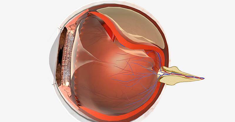
This period is one of the most critical phases of expected recovery, as the near‑final picture of retinal function becomes clearer, particularly if the macula (the central part of the retina responsible for sharp vision) was not severely damaged before surgery.
The patient may need an updated eyeglass prescription or low‑vision aids to optimize visual quality during these months after retinal detachment repair.
The ophthalmologist will continue to monitor retinal stability, watch for any traction or proliferative vitreoretinopathy, and in some cases determine the appropriate timing for silicone oil removal if it was used during surgery.
Long‑Term Stability (6 Months and Beyond)
Six months or more after retinal detachment surgery, most patients reach a relatively stable level of vision, with changes occurring more slowly and less noticeably than in the earlier months.
At this stage, long‑term success is mainly reflected by the retina remaining attached and the absence of recurrent detachment.
Some permanent effects may persist, such as partial reduction in visual acuity or loss of a portion of the visual field, especially in cases of extensive detachment or delayed surgical intervention.
Even so, most patients can adapt to their remaining level of vision and benefit from appropriate glasses or visual rehabilitation aids, allowing them to carry out daily activities in a way that is close to their normal routine.
Annual follow‑up with an ophthalmologist remains important after retinal detachment surgery to detect any new changes, protect the fellow eye, and manage risk factors such as high myopia or a family history of retinal disease.
What happens to the eye after surgery, and what affects temporary vision changes?
Following retinal detachment surgery, many patients notice blurred or reduced vision in the operated eye. This is expected and considered normal during the recovery period.
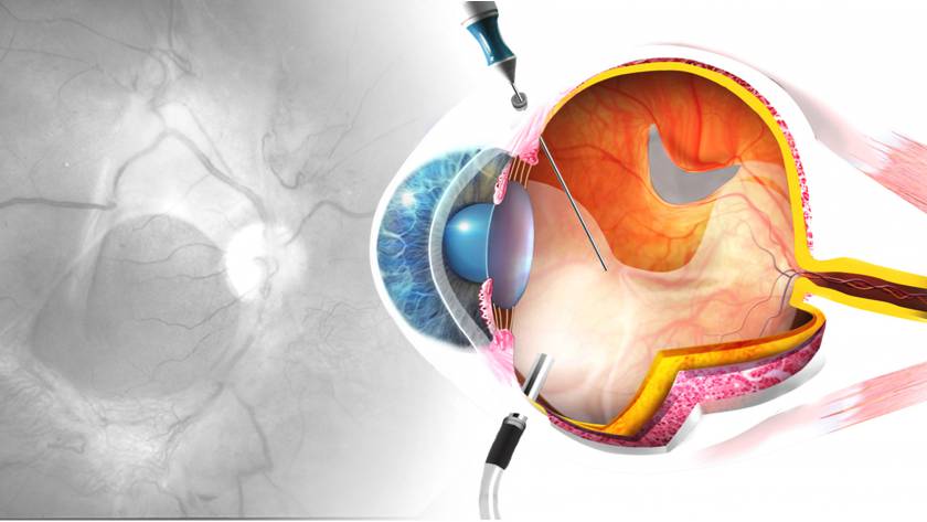
The main reasons are internal swelling in the eye, the presence of a gas bubble or silicone oil used to keep the retina in place, the effect of surgical sutures, and temporary changes in intraocular pressure.
You may also see floaters, flashes of light, or experience changes in colour perception and visual sharpness. These symptoms usually improve gradually as the retina heals and the gas bubble is absorbed, or after the silicone oil is removed at the time your doctor recommends.
The condition of the eye’s natural lens can also affect temporary visual quality, for example the early development of cataract, as well as dryness of the ocular surface or the use of certain postoperative eye drops.
Regular follow‑up with your ophthalmologist and strict adherence to post‑operative instructions after retinal detachment surgery help vision stabilise more quickly and reduce the risk of complications. Final visual recovery, however, can take several weeks to months, depending on the condition of the retina before surgery, the extent of the detachment, and the overall success of the procedure.
Factors Influencing the Speed and Extent of Visual Recovery (Aligned with the Study Objective)
The speed and extent of visual recovery after retinal detachment surgery are determined by several interrelated factors. The most critical are the involvement of the macula (the central area of sharp vision) and the duration of macular detachment before surgical intervention. The earlier the retina is reattached—preferably before the detachment reaches the macula—the better and faster the recovery of visual acuity tends to be.
The type of retinal detachment (tractional, rhegmatogenous, or exudative) and the surgical technique employed (such as intravitreal gas injection, silicone oil tamponade, cryotherapy, or laser photocoagulation) also play a key role in both the rate of visual improvement and the final level of visual clarity.
Visual outcomes are further affected by the patient’s age; the presence of systemic comorbidities such as diabetes mellitus and hypertension; and pre‑existing ocular conditions, including high myopia or a history of prior retinal or lens surgery.
Strict adherence to postoperative instructions—such as maintaining the prescribed head positioning, using eye drops as directed, and avoiding strenuous activity or prolonged bending—directly contributes to faster recovery and reduces the risk of complications that may delay visual improvement.
Ultimately, visual recovery after retinal detachment repair is gradual and may take from several weeks to several months. Final visual outcomes vary from one patient to another, depending on the preoperative status of the retina and the eye’s individual response to treatment.
When Is Recovery Considered Normal, and When Is It Concerning?

After retinal detachment surgery, recovery is generally considered normal if you notice a gradual reduction in visual haze and a slow improvement in visual acuity over the course of several weeks to months. It is also reassuring if bright flashes of light diminish, floaters become fewer without any new disturbing visual symptoms, and there is no severe eye pain, marked redness, or sudden onset headache.
On the other hand, recovery becomes worrisome if your vision stops improving altogether, starts to deteriorate after having been stable, or if you notice a dark curtain or shadow encroaching on your field of vision. A sudden increase in floaters or flashes of light, sharp pain inside the eye, pronounced eyelid swelling, or abnormal ocular discharge are also red-flag symptoms. These may indicate recurrent retinal detachment, elevated intraocular pressure, or intraocular infection (endophthalmitis), and require urgent evaluation by an ophthalmologist without waiting for your routine follow‑up appointment.
Vision‑Affecting Complications After Surgery
Following retinal detachment surgery, some patients may experience complications that temporarily or permanently affect visual quality, which makes close follow‑up with the ophthalmologist essential.
Blurred or hazy vision can persist for several weeks to months due to retinal edema or the natural slowness of tissue healing. This is common after retinal detachment repair and does not necessarily indicate surgical failure.
Other possible complications include recurrent retinal detachment, visual distortions (such as wavy or warped images), or loss of part of the visual field, especially if the detachment was extensive or long‑standing before surgery.
Visual acuity may also be reduced by cataract formation following surgery, particularly when intraocular gas or silicone oil has been used. In such cases, cataract extraction may later be required to restore and improve vision.
In some patients, epiretinal membranes may form on the surface of the retina, leading to image distortion or diplopia (double vision). When significant, these membranes can be surgically removed.
Regular postoperative follow‑up, adherence to medical instructions, and prompt reporting of any sudden drop in vision, new flashes of light, or a marked increase in floaters are crucial for detecting complications early and maximizing the chances of preserving the best possible vision.
Medical Tips to Speed Up Visual Recovery and Reduce Complications (Retinal Detachment Surgery)
To promote faster visual recovery after retinal detachment surgery and lower the risk of complications, ophthalmologists strongly recommend strict adherence to the postoperative instructions provided by your retinal surgeon.
Maintaining the prescribed head positioning is especially critical when an intraocular gas bubble has been used, as this specific posture helps keep the retina in place, enhances the likelihood of surgical success, and improves the chances of visual restoration.
Use your prescribed eye drops—such as antibiotics, anti‑inflammatory agents, and any other medications—exactly according to schedule. Do not stop them or adjust the doses without consulting your doctor, as proper use lowers the risk of infection and elevated intraocular pressure.
Avoid heavy lifting, sudden bending, strenuous exercise, or any activity that may increase intraocular pressure during the first few weeks after retinal detachment surgery. This precaution helps reduce the chances of redetachment or intraocular bleeding.
Protect the operated eye from trauma and friction, and avoid rubbing it or exposing it to dust and contaminated water. Wearing medical-grade sunglasses outdoors can also help reduce light sensitivity.
Quitting smoking, controlling chronic conditions such as diabetes and hypertension, maintaining good hydration, and following a diet rich in antioxidants and omega‑3 fatty acids (such as fatty fish and leafy green vegetables) all play a role in supporting retinal health after surgery.
Keep all follow‑up appointments as scheduled, and contact your ophthalmologist immediately if you notice sudden worsening of vision, a sudden increase in floaters, flashes of light, or severe eye pain. Prompt evaluation and management of these symptoms significantly reduce the risk of long-term complications following retinal detachment surgery.
Follow‑up Schedule and Tests to Assess Visual Recovery
After retinal detachment surgery, the ophthalmologist will schedule regular follow‑up visits to monitor visual recovery and ensure the retina remains properly attached.
The first visit is usually within the first week after surgery, followed by several appointments over the next few weeks. As the eye stabilizes and vision improves, the interval between visits is gradually increased, depending on your clinical progress.
At each follow‑up, your doctor will typically check visual acuity, measure intraocular pressure, and perform a dilated fundus examination to confirm that the retina is healing and that there are no new areas of fluid leakage or bleeding. When indicated, additional tests may be ordered, such as optical coherence tomography (OCT) or fluorescein angiography.
Keeping these appointments is essential for early detection of complications such as recurrent retinal detachment or elevated intraocular pressure, and for assessing the degree of visual improvement. Based on the findings, your doctor may recommend further treatments or tailored visual rehabilitation exercises.
Visual recovery after retinal detachment surgery is often gradual and varies from one patient to another, so the final outcome should not be judged before several months have passed and all recommended follow‑up evaluations have been completed.
Best Retina Specialist in Jeddah for Postoperative Care After Retinal Detachment Surgery – Dr. Waddah Jalabi (Search-Intent Optimized)
Ensuring follow-up with the best retina specialist in Jeddah after retinal detachment surgery is crucial for the success of the procedure and for preserving vision in the long term. Dr. Waddah Jalabi is considered one of the leading names in this field, thanks to his extensive experience in vitreoretinal surgery and his meticulous approach to postoperative care.
Dr. Waddah Jalabi is committed to developing an individualized follow-up plan for each patient after retinal detachment repair. This typically includes scheduled visits for dilated fundus examination, monitoring retinal stability, measuring intraocular pressure, and early detection of any complications such as recurrent retinal detachment, vitreous hemorrhage, or ocular hypertension.
He also provides clear, simplified explanations of all post-op instructions, including head positioning, proper use of eye drops, and the safe timing for returning to daily activities. This helps patients understand their condition better and significantly reduces anxiety.
Choosing an ophthalmologist who specializes in post-retinal detachment follow-up, such as Dr. Waddah Jalabi in Jeddah, not only improves surgical outcomes, but also maximizes the chances of preserving as much vision as possible, while ensuring safe, evidence-based medical care aligned with the latest clinical guidelines.
Book Your Post-Retinal Detachment Surgery Follow-Up at Batal Specialist Eye Center
Schedule your follow-up visit now at Batal Specialist Eye Center to ensure a safe recovery after retinal detachment surgery and to closely monitor the stability of your vision, step by step.
Our consultant retinal specialists are committed to:
- Assessing visual acuity
- Controlling intraocular inflammation and eye pressure
- Confirming that the retina remains attached
- Detecting any early signs of complications, such as recurrent detachment or vitreous hemorrhage
At Batal Specialist Eye Center, our dedicated follow-up clinics offer flexible appointment times and advanced diagnostic imaging, including Optical Coherence Tomography (OCT) of the retina and fundus photography. These tools help us detect and treat any problem at an early stage—before it can affect your visual quality.
Do not wait for symptoms to reappear. Regular follow-up after retinal detachment surgery is a crucial part of long-term surgical success and essential to protecting your eye from permanent vision loss.
Book your follow-up appointment now—by phone or online—with Batal Specialist Eye Center and gain reassurance about the health of your retina and the stability of your vision.


