
Med info
Age-Related Macular Degeneration: Symptoms and the Difference Between Dry and Wet AMD

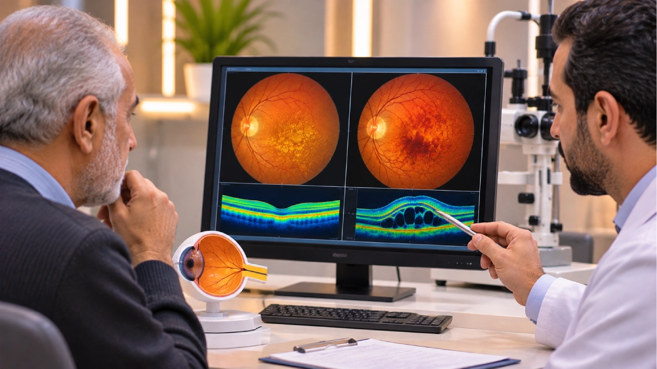
Vision changes can begin subtly. Straight lines may appear wavy, words may become harder to read, or recognizing familiar faces may require more effort. While these changes are sometimes mistaken for a normal part of aging, they may indicate a condition affecting the central part of the retina.
Age-related macular degeneration (AMD) is an eye disease that affects the macula, the part of the retina responsible for sharp, detailed central vision. It has two main forms: dry AMD and wet AMD.
Dry AMD usually progresses gradually, while wet AMD can cause more rapid vision deterioration due to the growth of abnormal blood vessels that leak fluid or blood into the retina.
Early detection helps determine the type and stage of the disease, allowing ophthalmologists to recommend appropriate monitoring or treatment to reduce the risk of further vision loss.
What Is Age-Related Macular Degeneration?
Age-related macular degeneration is a retinal disease that affects the macula, a small area near the center of the retina responsible for detailed vision.
The macula plays an essential role in reading, recognizing faces, driving, distinguishing colors, and performing activities that require precise visual focus.
When the macula becomes damaged, central vision may appear blurred or distorted, even when peripheral vision remains relatively unaffected.
AMD does not usually cause complete blindness. However, advanced disease can lead to severe central vision impairment, affecting independence and everyday activities.
To understand how AMD affects sight, it is helpful to know how the retina receives light and converts it into nerve signals. The macula is the specialized part of the retina responsible for fine visual detail, which explains why AMD symptoms differ from those of retinal diseases affecting other areas of vision.

What Is the Difference Between Dry and Wet Macular Degeneration?
Dry and wet AMD differ in the changes they cause within the retina, how quickly they progress, and the treatment options available.
Dry Age-Related Macular Degeneration (Dry AMD)
Dry AMD is the more common form of age-related macular degeneration.
It develops when the tissues of the macula undergo gradual degenerative changes associated with aging.
Small deposits called drusen may accumulate beneath the retina, alongside changes in the cells that support normal visual function.
Dry AMD is generally classified into early, intermediate, and advanced stages.
During the early stages, patients may experience few or no noticeable symptoms.
In advanced dry AMD, a condition known as geographic atrophy may develop. This involves the progressive loss of retinal cells in specific areas of the macula.
Wet Age-Related Macular Degeneration (Wet AMD)
Wet AMD, also known as neovascular AMD, occurs when abnormal blood vessels develop beneath or within the retina.
These vessels may leak fluid or blood, disrupting the structure of the macula and damaging central vision.
Although wet AMD is less common than dry AMD, it can progress more rapidly and requires prompt specialist assessment and treatment.
Dry AMD can progress to wet AMD, making regular monitoring important even when vision appears stable.
Dry AMD vs. Wet AMD: Key Differences
| Feature | Dry AMD | Wet AMD |
| Underlying changes | Progressive degeneration and thinning of macular tissue | Growth of abnormal blood vessels |
| Prevalence | More common | Less common |
| Progression | Usually gradual | May be rapid |
| Symptoms | Blurred central vision and difficulty seeing details | Distorted lines or rapid central vision deterioration |
| Treatment | Monitoring, risk reduction, and selected stage-specific treatments | Primarily anti-VEGF injections |
| Disease course | May progress to wet AMD | Represents the neovascular form of advanced AMD |
Symptoms alone cannot reliably distinguish dry AMD from wet AMD.
An accurate diagnosis requires a retinal examination and appropriate imaging.
What Are the Symptoms of Age-Related Macular Degeneration?
AMD may not cause noticeable symptoms during its early stages, particularly in patients with dry AMD.
As the disease progresses, changes affecting central vision may become more apparent.
Common symptoms include:
- Blurred vision in the center of the visual field.
- Straight lines appearing wavy, bent, or distorted.
- Difficulty reading small print.
- Needing brighter lighting when reading.
- Difficulty recognizing faces.
- A blurred or dark spot in central vision.
- Reduced clarity of colors and fine details.
One eye may become affected before the other, making early changes harder to recognize because the healthier eye can partially compensate for reduced vision.
Are Wavy or Distorted Straight Lines a Serious Warning Sign?
Seeing straight lines as wavy or distorted may indicate changes in the macula, including those associated with wet AMD.
If this symptom develops suddenly or is accompanied by a rapid decline in central vision, seek urgent ophthalmic evaluation, preferably on the same day.
Do not wait for the symptoms to resolve on their own.
Delaying the assessment of certain macular conditions may reduce the opportunity to preserve vision.
What Causes Age-Related Macular Degeneration?
There is no single cause responsible for every case of AMD.
The disease is associated with age-related changes in the retina, along with genetic and environmental factors.
Important risk factors include:
Age: The risk of AMD increases after the age of 55 and continues to rise with advancing age.
Family history: People with relatives who have AMD may face a higher risk of developing the condition.
Smoking: Smoking is a significant modifiable risk factor associated with both AMD development and disease progression.
Cardiovascular health: Certain conditions affecting blood vessel health may contribute to the risk or progression of AMD.
Having these risk factors does not mean that AMD will necessarily develop.
However, they can help an ophthalmologist determine whether regular retinal examinations are recommended.
How Is Age-Related Macular Degeneration Diagnosed?
Diagnosing AMD involves evaluating visual function, examining the retina, and using specialized imaging to identify changes in the macula.
Dilated Retinal Examination
During a dilated eye examination, the ophthalmologist assesses the retina and macula for drusen, pigment changes, fluid accumulation, or bleeding.
This examination may reveal early signs of AMD before patients notice significant changes in vision.
Optical Coherence Tomography (OCT)
Optical Coherence Tomography (OCT) is one of the most important imaging techniques used to evaluate macular diseases.
It produces detailed cross-sectional images of the retina, allowing ophthalmologists to identify structural abnormalities, fluid accumulation, and signs of retinal atrophy.
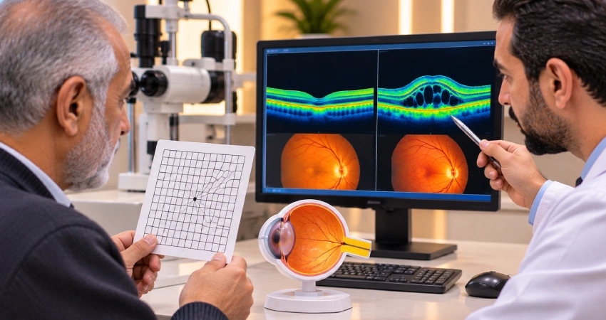
OCT is particularly valuable when wet AMD is suspected and is also used to monitor the response to treatment.
Retinal Angiography
Some patients may require fluorescein angiography or OCT angiography (OCT-A) to evaluate abnormal retinal blood vessels.
The choice of imaging depends on the findings of the initial examination and the ophthalmologist’s clinical assessment.
These tests also help distinguish AMD from other conditions that affect central vision.
For example, diabetic retinopathy can cause fluid leakage and swelling in the macula, but its underlying mechanisms and treatment approaches differ from those of age-related macular degeneration.
Can Dry Age-Related Macular Degeneration Be Treated?
Management of dry AMD depends on the stage of the disease and the degree of visual impairment.
During the early stages, an ophthalmologist may recommend regular monitoring and lifestyle changes to address modifiable risk factors.
These may include stopping smoking, maintaining physical activity, and following a balanced diet.
For certain patients with intermediate AMD, nutritional supplements containing the AREDS2 formulation may help reduce the risk of progression to advanced disease.
However, AREDS2 supplements are not suitable for every patient and cannot restore vision that has already been lost.
They should only be used after an ophthalmologist has assessed the patient’s condition and determined whether they are appropriate.
Are Treatments Available for Geographic Atrophy?
In some countries, intravitreal medications targeting components of the complement system are available to slow the enlargement of geographic atrophy lesions in selected patients.
These treatments do not restore damaged retinal cells or guarantee improved vision.
They also carry potential risks that should be discussed with a retina specialist.
Availability and regulatory approval vary between countries, so treatment decisions must take into account the patient’s condition and the therapies approved and accessible locally.
How Is Wet Age-Related Macular Degeneration Treated?
Intravitreal injections of anti-vascular endothelial growth factor (anti-VEGF) medications are a primary treatment for many patients with wet AMD.
These medications help suppress abnormal blood vessel activity and reduce fluid leakage within the retina.
Treatment may stabilize vision or improve it in some patients.
Are Eye Injections for Wet AMD Painful?
Intravitreal injections are generally performed after applying local anesthetic eye drops and disinfecting the eye.
Patients may experience mild pressure or brief discomfort during the procedure.
The ophthalmologist determines the number of injections and the intervals between treatments based on retinal response and OCT findings.
Some patients require repeated injections over an extended period because the disease can become active again after an initial improvement.
Intravitreal injections are also used to treat other retinal conditions, including diabetic macular edema. However, the medication and follow-up schedule depend on the underlying diagnosis.
Can Wet AMD Be Treated with Laser Therapy?
Photodynamic therapy (PDT) may be considered in selected cases, sometimes alongside intravitreal injections.
However, it is not the usual first-line treatment for most patients with wet AMD.
The decision depends on the characteristics of the abnormal blood vessels and the eye’s response to treatment.
Can Vision Be Restored After Macular Degeneration?
The possibility of visual improvement depends on the type and stage of AMD and the extent of retinal damage.
In wet AMD, anti-VEGF injections may improve vision in some patients, particularly when treatment begins early.
However, treatment cannot guarantee complete visual recovery.
Advanced damage caused by the loss of macular cells is generally irreversible with currently available treatments.
For this reason, ongoing care focuses on detecting changes early, slowing disease progression, and using low-vision rehabilitation when appropriate.
How Can Patients Monitor Macular Degeneration at Home?
An ophthalmologist may recommend using an Amsler grid, a simple chart of straight lines that can help patients notice certain changes in central vision.
To use the grid:
- Wear your usual reading glasses if needed.
- Cover one eye.
- Look directly at the central dot.
- Check whether any lines appear wavy, distorted, or missing.
- Repeat the test with the other eye.
If you notice new distortion or missing areas, contact your ophthalmologist promptly.
An Amsler grid cannot detect every case of AMD and does not replace retinal examinations or OCT imaging.
Can Age-Related Macular Degeneration Be Prevented?
There is no guaranteed way to prevent AMD.
However, certain measures may help reduce modifiable risk factors and support overall eye health.
These include:
- Avoiding smoking.
- Maintaining regular physical activity.
- Eating a balanced diet that includes leafy green vegetables and fish.
- Managing blood pressure, cholesterol, and chronic health conditions.
- Attending regular eye examinations when risk factors are present.
- Avoiding AREDS2 supplements unless they are appropriate for the stage of AMD.
Specialist monitoring remains important because early retinal changes may develop without noticeable symptoms.
Macular Disease Diagnosis and Follow-Up with Dr. Waddah Chalabi
Macular diseases require careful retinal assessment, particularly when patients develop distorted vision or sudden changes in central visual clarity.
Dr. Waddah Chalabi is a Consultant Vitreoretinal Surgeon and Medical Director at Batal Eye Center, with more than 30 years of professional experience in diagnosing and treating complex retinal conditions.
He holds a Fellowship of the Royal College of Surgeons (FRCS).
His expertise includes advanced retinal surgery, diabetic retinopathy management, and the evaluation of retinal diseases using OCT imaging and retinal photography.
Specialist experience supports the assessment of macular changes, helps distinguish between different causes of central vision impairment, and guides decisions regarding monitoring or treatment.
Experiencing Blurred Central Vision? Book a Retinal Examination at Batal Eye Center
Age-related macular degeneration may begin with subtle changes in reading or the ability to distinguish fine details.
Some forms can progress rapidly, particularly when abnormal blood vessels develop beneath the retina.
Batal Eye Center in Jeddah provides retinal evaluation services, including dilated retinal examinations and OCT imaging when clinically indicated.
These assessments help identify the cause of visual changes and determine the most appropriate management plan.
If you notice newly distorted straight lines, a dark spot in the center of your vision, or a rapid decline in visual clarity, do not delay medical evaluation.
Frequently Asked Questions About Age-Related Macular Degeneration
What Is the Difference Between Dry and Wet AMD?
Dry AMD involves gradual degenerative changes in macular tissue.
Wet AMD develops when abnormal blood vessels grow beneath or within the retina and may leak fluid or blood, potentially causing more rapid deterioration of central vision.
Can Macular Degeneration Cause Complete Blindness?
AMD does not usually cause complete blindness.
However, it can result in severe central vision loss, affecting reading, facial recognition, and daily activities.
Can Dry AMD Turn into Wet AMD?
Yes. Dry AMD can progress to wet AMD.
Regular monitoring and prompt evaluation of new visual changes are therefore important.
What Are the First Signs of Macular Degeneration?
Early AMD may cause no noticeable symptoms.
As the disease progresses, patients may experience blurred central vision, difficulty reading, or straight lines appearing wavy.
Can Macular Degeneration Be Completely Cured?
There is currently no cure that restores all retinal tissue damaged by AMD.
However, available treatments may slow the progression of certain forms of the disease or help stabilize vision, particularly in wet AMD.
Do Eye Injections Treat Wet Macular Degeneration?
Anti-VEGF injections are a primary treatment for many patients with wet AMD.
They can reduce fluid leakage and may help preserve or improve vision.
Is OCT Necessary for Patients with AMD?
OCT is widely used to evaluate the macula, detect fluid accumulation and structural changes, and monitor disease progression or treatment response.
Is Age-Related Macular Degeneration Hereditary?
Genetic factors can increase the risk of AMD, but the condition is not necessarily passed directly from parents to children.
Age, smoking, and other factors also contribute to the likelihood of developing the disease.
Can Vitamins Treat Macular Degeneration?
AREDS2 supplements may reduce the risk of disease progression in certain patients.
However, they are not effective for every stage of AMD and cannot restore lost vision.
An ophthalmologist should determine whether supplementation is appropriate.
When Should I See a Retina Specialist Urgently?
Seek urgent ophthalmic assessment if you develop newly distorted straight lines, a sudden dark spot in central vision, or a rapid decline in visual clarity.