
Med info
Diabetic Macular Edema: Symptoms, Treatment Options, and Follow-Up Care

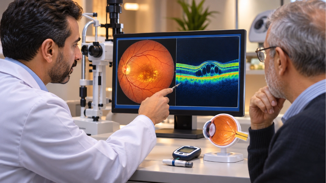
Vision changes in people with diabetes often develop gradually. Words may become difficult to read, straight lines may appear wavy, or fine details may become harder to distinguish. In some cases, these symptoms result from fluid accumulating in the central part of the retina.
Diabetic macular edema (DME) is a diabetes-related eye complication that occurs when damaged retinal blood vessels leak fluid into the macula, the area responsible for sharp central vision. This swelling can cause blurred or distorted vision, although some patients experience no noticeable symptoms during the early stages.
Early diagnosis through a comprehensive retinal examination and Optical Coherence Tomography (OCT) helps determine the extent of swelling and guides appropriate treatment or monitoring to protect vision.
What Is Diabetic Macular Edema?
Diabetic macular edema is a complication of diabetes that affects the retina when fluid leaks from damaged small blood vessels into the macular tissue.
The macula is located at the center of the retina and is responsible for the detailed vision needed for reading, recognizing faces, driving, and other everyday activities.
When fluid accumulates in this area, it can interfere with the normal function of light-sensitive retinal cells, resulting in blurred or distorted central vision.
DME may affect one or both eyes and can develop at different stages of diabetic retinopathy.
Although the two conditions are closely related, they are not identical. Diabetic retinopathy refers to a broader range of diabetes-related changes affecting retinal blood vessels, whereas diabetic macular edema specifically involves fluid accumulation in the macula.
Understanding the symptoms and treatment of diabetic retinopathy can help explain how diabetes affects retinal blood vessels and why macular swelling requires careful evaluation.

What Causes Diabetic Macular Edema?
Diabetic macular edema develops primarily because prolonged high blood sugar levels can damage the small blood vessels supplying the retina.
Over time, the walls of these vessels may weaken and become more permeable, allowing fluid to leak into the surrounding retinal tissue.
Several factors may increase the likelihood of developing DME or contribute to its progression:
- Poor long-term blood sugar control.
- High blood pressure.
- Abnormal cholesterol and blood lipid levels.
- Longer duration of diabetes.
- Existing diabetic retinopathy.
- Certain diabetes-related kidney conditions.
Having one or more of these risk factors does not necessarily mean that macular edema will develop.
However, they highlight the importance of regular retinal examinations and coordinated care between an ophthalmologist and the physician managing diabetes.
What Are the Symptoms of Diabetic Macular Edema?
Diabetic macular edema may not cause noticeable symptoms in its early stages, particularly when swelling is mild or does not significantly affect the center of the macula.
As the condition progresses, patients may experience:
- Blurred central vision.
- Difficulty reading small print.
- Straight lines appearing bent or wavy.
- Reduced ability to distinguish fine details.
- Changes in color perception.
- A blurred or hazy area in the center of vision.
Symptoms may develop gradually. However, sudden vision loss requires urgent medical evaluation, as it may indicate another serious eye complication, such as vitreous hemorrhage or an acute retinal disorder.
Can Diabetic Macular Edema Occur Without Noticeable Vision Loss?
Yes. OCT imaging may detect fluid accumulation in the macula before a patient notices any significant change in vision.
For this reason, the absence of symptoms does not necessarily indicate that the retina is healthy.
Regular retinal screening remains important for people with diabetes, even when their vision appears normal.

How Is Diabetic Macular Edema Diagnosed?
Diagnosing DME involves evaluating visual acuity, examining the retina, and using specialized imaging to determine where fluid has accumulated and whether the center of the macula is affected.
Dilated Retinal Examination
During a retinal examination, the ophthalmologist assesses the blood vessels and surrounding retinal tissue, often after dilating the pupils.
The examination may reveal signs of diabetic retinopathy, including small retinal hemorrhages, hard exudates, and other vascular abnormalities.
Although a dilated examination provides valuable information about retinal health, additional imaging may be necessary to accurately assess macular swelling.
Optical Coherence Tomography (OCT)
OCT is one of the most important diagnostic and monitoring tools for diabetic macular edema.
This non-invasive imaging technique produces detailed cross-sectional images of the retina, allowing the ophthalmologist to:
- Identify fluid accumulation within retinal layers.
- Measure macular thickness.
- Assess the location and extent of swelling.
- Determine whether the edema involves the center of the macula.
- Compare findings across follow-up appointments.
Determining whether DME affects the central macula is particularly important because it influences treatment decisions.
Fluorescein Angiography
In selected cases, an ophthalmologist may recommend fluorescein angiography to identify areas of vascular leakage or evaluate abnormalities in retinal blood circulation.
This test involves using a fluorescent dye to visualize retinal blood vessels.
Not every patient requires fluorescein angiography. Its use depends on the clinical examination, OCT findings, and the potential treatment plan.
Together, these investigations help distinguish the broader vascular changes of diabetic retinopathy from the specific impact of fluid leakage on central vision.
Does Every Patient with Diabetic Macular Edema Need Immediate Treatment?
Not every case of diabetic macular edema requires immediate injections or laser treatment.
The decision depends on the location of the swelling, the patient’s visual acuity, and the findings of OCT imaging.
For some patients with center-involving DME who still have good visual acuity, careful observation may be an appropriate initial approach.
A clinical study conducted by the DRCR Retina Network found that structured observation, with treatment initiated if vision declined, was a reasonable strategy for certain patients with center-involving DME and good baseline vision.
However, when macular swelling affects central vision and causes visual impairment, intravitreal injections are commonly considered a primary treatment option.
The decision to move from monitoring to active treatment depends on the patient’s actual retinal findings rather than the presence of swelling alone.
This distinction is also explored in the article on when diabetic retinopathy requires treatment rather than monitoring, which discusses how disease progression and clinical findings influence management decisions.
What Are the Treatment Options for Diabetic Macular Edema?
Several treatments are available for diabetic macular edema.
A retina specialist selects the most appropriate approach based on the location and severity of swelling, visual acuity, previous treatments, and the patient’s response.
1. Anti-VEGF Eye Injections
Intravitreal anti-vascular endothelial growth factor (anti-VEGF) injections are a major treatment option for center-involving diabetic macular edema associated with reduced vision.
These medications work by blocking vascular endothelial growth factor, a protein involved in increased blood vessel permeability.
By reducing vascular leakage, anti-VEGF treatment can help decrease retinal swelling and improve or stabilize vision.
Medications used in selected cases include:
- Aflibercept.
- Ranibizumab.
- Faricimab.
The choice of medication depends on local regulatory approval, clinical findings, and the treating specialist’s assessment.
Although many patients benefit from anti-VEGF therapy, responses vary. Treatment cannot guarantee complete recovery of vision, particularly when longstanding retinal damage is present.
2. Focal Laser Treatment
Focal laser treatment may be considered for certain cases of diabetic macular edema, particularly when the swelling does not involve the center of the macula.
The procedure targets specific areas of vascular leakage to help reduce the risk of further retinal damage.
It differs from the laser treatment used for abnormal blood vessel growth in proliferative diabetic retinopathy.
Focal laser treatment is not automatically a substitute for anti-VEGF injections when center-involving DME causes reduced vision.
3. Intravitreal Corticosteroid Treatment
Corticosteroid injections or implants may be considered for selected patients, including some individuals who have not responded adequately to initial treatment.
These medications help reduce inflammation and vascular leakage.
However, corticosteroid treatment may increase intraocular pressure or accelerate cataract development.
For this reason, ophthalmologists carefully evaluate the potential benefits and risks and monitor eye pressure during treatment.
4. Managing Diabetes and Associated Health Conditions
Effective management of blood sugar, blood pressure, and cholesterol is an essential part of caring for patients with diabetic macular edema.
Good metabolic and cardiovascular risk-factor control may help reduce the likelihood of further retinal vascular damage.
However, controlling blood sugar alone does not replace ophthalmic treatment when existing macular edema requires intervention.
The most appropriate treatment plan combines retinal care with management of the underlying systemic conditions.
Anti-VEGF Injections vs. Laser Treatment for Diabetic Macular Edema
| Feature | Anti-VEGF Injections | Focal Laser Treatment |
| Main purpose | Reduce vascular leakage and macular swelling | Treat specific areas of leakage |
| Common indications | Many cases of center-involving DME with reduced vision | Selected cases of non-center-involving DME |
| Treatment frequency | Repeated injections may be necessary | Depends on the condition and response |
| Visual outcomes | Vision may improve or stabilize | Often aims to reduce the risk of deterioration |
| Monitoring | Visual acuity assessment and OCT | Retinal examination and OCT when indicated |
Some patients may require more than one treatment approach, particularly when the response to initial therapy is insufficient.
How Many Eye Injections Are Needed for Diabetic Macular Edema?
There is no fixed number of injections suitable for every patient.
Some treatment regimens begin with relatively frequent injections, often at approximately monthly intervals, followed by adjustments based on visual improvement and OCT findings.
Certain patients require ongoing treatment over an extended period, while others may gradually have longer intervals between injections once their condition stabilizes.
Needing additional injections does not necessarily mean that treatment has failed.
DME can be chronic or recurrent, making continued monitoring and repeated treatment necessary for some patients.
Following the recommended injection schedule is important because stopping treatment without medical assessment may allow fluid to accumulate again and vision to deteriorate.
How Long Does It Take for Vision to Improve After DME Treatment?
Some patients notice visual improvement within the first few weeks or months of treatment, while others may require more time.
The outcome depends on several factors, including:
- How long the macular edema has been present.
- The severity of retinal swelling.
- The condition of the macular tissue.
- The extent of existing retinal damage.
- The patient’s response to treatment.
OCT imaging may show a reduction in retinal thickness without an equivalent improvement in visual acuity.
This can occur when prolonged disease has already affected the retinal cells responsible for detailed vision.
Treatment success is therefore evaluated through both anatomical improvements and changes in visual function.
Can Diabetic Macular Edema Return After Treatment?
Yes. Diabetic macular edema can recur after an initial improvement.
Fluid may accumulate again if the underlying vascular abnormalities persist or the effects of treatment diminish over time.
Recurrence can occur even in patients who previously responded well to injections.
Regular OCT monitoring helps detect renewed swelling and allows the ophthalmologist to adjust the treatment plan when necessary.
Maintaining blood sugar, blood pressure, and cholesterol levels within the targets recommended by the treating physician is also important for reducing the risk of diabetes-related eye complications.
Can Diabetic Macular Edema Be Prevented?
Not every case of diabetic macular edema can be prevented. However, appropriate diabetes management and regular eye care can help reduce the risk of developing the condition or experiencing further progression.
Important preventive measures include:
- Maintaining blood sugar levels according to the prescribed diabetes management plan.
- Monitoring blood pressure and cholesterol.
- Attending regular retinal examinations, even without symptoms.
- Avoiding smoking.
- Seeking ophthalmic evaluation when new vision changes occur.
- Following prescribed injection and monitoring schedules after a DME diagnosis.
Regular screening is especially important because retinal vascular damage and macular swelling may develop before significant visual impairment becomes apparent.
The importance of early diabetic retinopathy assessment and treatment lies in identifying these changes and determining when specialized investigations or treatment may be necessary.
Diabetic Macular Edema Diagnosis and Treatment with Dr. Waddah Chalabi
Managing diabetic macular edema requires specialized assessment to determine the location and severity of retinal swelling, evaluate its effect on central vision, and select an appropriate treatment strategy.
Dr. Waddah Chalabi is a Consultant Vitreoretinal Surgeon and Medical Director at Batal Eye Center, with more than 30 years of professional experience in retinal diseases and surgery.
He holds a Fellowship of the Royal College of Surgeons (FRCS), and his clinical expertise includes diabetic retinopathy management, advanced retinal surgery, and the assessment of retinal diseases using specialized diagnostic imaging.
Specialist evaluation helps distinguish cases that can be monitored from those requiring intravitreal injections, laser treatment, or other interventions.
It also supports ongoing assessment of retinal response to treatment and appropriate adjustments to the management plan
Experiencing Blurred Vision with Diabetes? Book a Retinal Examination at Batal Eye Center
Diabetic macular edema may develop without obvious symptoms in its early stages.
However, persistent fluid accumulation can gradually affect central vision, making reading and other daily activities more difficult.
At Batal Eye Center in Jeddah, patients can access diagnostic and management services for diabetes-related retinal conditions, including retinal examinations and OCT imaging.
These assessments help determine whether the macula is affected and whether monitoring or treatment is appropriate.
If you have diabetes and are experiencing blurred vision, or previous examinations have identified retinal swelling or fluid leakage, an assessment by a retina specialist can help determine the next step.
Contact Batal Eye Center to schedule a retinal examination and discuss your diagnostic findings and available treatment options.
Frequently Asked Questions About Diabetic Macular Edema
Is Diabetic Macular Edema Serious?
Diabetic macular edema can cause permanent central vision impairment if swelling persists and retinal tissue becomes damaged.
Early diagnosis and appropriate management can help reduce the risk of further vision loss.
Is Diabetic Macular Edema the Same as Diabetic Retinopathy?
No. Diabetic retinopathy involves broader diabetes-related changes in retinal blood vessels, while diabetic macular edema specifically refers to fluid accumulation in the macula.
DME can develop at different stages of diabetic retinopathy.
Can Diabetic Macular Edema Be Completely Cured?
Macular swelling may improve or resolve in some patients following treatment.
However, the condition can recur, and certain patients require long-term treatment or monitoring.
Can Eye Injections Restore Vision?
Anti-VEGF injections may improve vision in some patients by reducing macular swelling.
However, complete visual recovery cannot be guaranteed, especially when chronic retinal damage has already occurred.
Does Every Patient with DME Need Eye Injections?
No. Structured observation may be appropriate for selected patients whose vision remains good.
Other patients may require injections or laser treatment depending on the location and severity of the edema.
How Often Should OCT Be Performed for Diabetic Macular Edema?
The frequency of OCT testing depends on the severity of swelling and the treatment plan.
OCT may be performed during injection appointments or follow-up visits to assess retinal response and determine whether additional treatment is needed.
Can Blood Sugar Control Alone Treat Diabetic Macular Edema?
Good blood sugar control is essential for reducing the risk of diabetic retinal complications.
However, it may not be sufficient to resolve existing macular edema, particularly when central vision is affected.
Can Diabetic Macular Edema Affect Only One Eye?
Yes. DME may affect one eye or both eyes, and the severity can differ between them.
Can Laser Treatment Replace Eye Injections?
Laser treatment may be suitable for selected cases, particularly when swelling does not involve the center of the macula.
For center-involving DME associated with reduced vision, anti-VEGF injections are commonly used as a primary treatment.
When Should I See a Retina Specialist Urgently?
Seek urgent ophthalmic evaluation if you experience sudden vision loss, a new dark spot, a sudden increase in floaters, or a curtain-like shadow covering part of your vision.
These symptoms may indicate a serious retinal complication requiring prompt assessment.