

Med info
Eyelid Lesions: Types, Symptoms, and Treatment Options
The skin of the eyelid is the thinnest, most exposed area of the body’s skin and contains nearly all cutaneous structures. This explains why the eyelids are particularly prone to developing benign, malignant, or cancerous lesions. Eyelid lesions consist of damaged or diseased cells that look noticeably different from the surrounding tissue.
All eyelid lesions arise from conditions affecting the different eyelid tissues, including the skin, fat, muscles, blood and lymphatic vessels, eyelashes, and more. Eyelid lesions are very common, and most cases resolve with minimal medical intervention, often through the topical application of antibiotic ointments to the lesion.
These lesions are frequently caused by infectious or pathological processes. However, some eyelid lesions require specialized, in‑depth medical care because they originate from malignant or cancerous tumors. It is therefore crucial to receive appropriate treatment for any type of lesion, as they are also cosmetically bothersome. For accurate diagnosis and effective treatment options, we recommend the Eye Center at Batal Specialized Complex in Saudi Arabia.
Also read: Thyroid Nodules | Causes, Symptoms, and Treatment of Thyroid Nodules
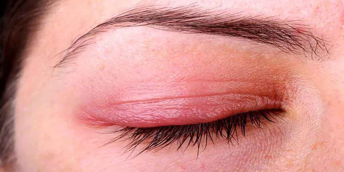
What Are the Early Symptoms of Eyelid Lesions?
Below are some visible signs of eyelid lesions that you should watch for, including:
[Here, bullet points or specific signs would normally be listed if provided in the original text.]
While these signs of eyelid lesions typically appear on the outer surface of the eyelid, they can also develop underneath it. In such cases, proper identification and treatment by a healthcare professional are essential.
Also read: Graves’ Disease | Everything You Need to Know About the Most Common Cause of Hyperthyroidism
Specific Causes and Symptoms of Benign Eyelid Lesions
Benign eyelid lesions are caused by pathological or physiological factors affecting eyelid tissues. These factors may include viral or bacterial infections or metabolic disorders. They include the following conditions:
Meibomian glands, located within the upper and lower eyelids, secrete an oily substance that helps keep the eye surface lubricated. When the Meibomian gland ducts become blocked by bacteria, the glandular secretions gradually accumulate. The result is a characteristic, painless, slow-growing lump on the upper or lower eyelid (commonly known as a chalazion).
Also read: Hyphema (Bleeding in the Eye) | Causes, Symptoms, and Treatment
Maintaining proper eyelid hygiene is effective in preventing chalazia. Treatment options for these eyelid lesions include topical and oral antibiotics, and if the chalazion persists, surgical excision of the lump may be required.
Xanthelasma
This type of eyelid lesion, called xanthelasma, occurs due to the buildup of lipid‑laden macrophages—immune system cells—within the eyelid skin. The increased presence of these macrophages in eyelid tissue is mainly related to underlying conditions such as lipid metabolism disorders, and sometimes chronic inflammatory or infectious processes. The result is a characteristic yellowish plaque on the upper and/or lower eyelid.
There is no fully reliable way to prevent xanthelasma. Treatment options include topical application of trichloroacetic acid (TCA), CO₂ laser therapy, and surgical excision of the lesion.
Epidermoid Cyst
An epidermoid (epidermal inclusion) cyst develops when an eyelash follicle becomes obstructed, often as a result of mechanical or blunt trauma. This blockage is preceded by the formation of a benign growth within the hair follicle, which then appears as a smooth, white, slow‑growing lesion along the eyelid margin.
There is no practical method to prevent epidermoid cysts. The primary treatment option for this condition is surgical removal of the benign cyst.
Also read: Cosmetic Botox Around the Eyes | Duration, Procedure, and Key Details
Apocrine Hidrocystoma
These eyelid lesions are caused by blockage of the apocrine sweat glands (such as the glands of Moll) located at the base of the eyelashes. The obstruction leads to accumulation of sweat within the gland, resulting in a smooth, round, translucent cystic lesion along the upper or lower eyelid margin, known as an apocrine hidrocystoma.
Regular eyelid and eye hygiene is an effective way to help prevent apocrine hidrocystomas. The main treatment for this condition is surgical incision and drainage or excision of the lesion.
Also read: Eye Massage | Detailed Information About Eye Massage
Nevi (Nevus of the Eyelid)
A nevus is a congenital abnormality that leads to the development of a tumor from undifferentiated melanocytes—the pigment‑producing cells—on the eyelids. This lesion appears as a small, well‑defined, light‑colored or pigmented bump on the upper or lower eyelid. While most nevi are benign, they require monitoring because some may undergo malignant transformation.
There is no effective way to prevent eyelid nevi. Treatment options, when indicated, include surgical excision of the lesion or wedge resection of the involved eyelid segment.
Also read: Healthy Diet | Everything You Need to Know and Its Impact on Eye Health
Specific Causes and Symptoms of Malignant Eyelid Lesions
Malignant eyelid lesions are associated with significant health risks, including a high likelihood of recurrence and a strong tendency to spread (metastasize) and cause cancerous growths in other parts of the body. Common malignant eyelid lesions seen in clinical practice include:
Actinic Keratosis
Actinic keratosis is caused by chronic sun‑induced skin damage affecting the upper or lower eyelid. It is characterized by a firm, rough, pink‑to‑red plaque on the eyelid surface. In more severe cases, the skin around the thickened plaque may become inflamed and can sometimes bleed.
Eyelid lesions due to actinic keratosis can be prevented by the regular use of sunscreen to protect the skin from ultraviolet (UV) damage. Treatment options include microscopic surgical excision of the lesion, along with topical chemotherapeutic agents. Cryotherapy (freezing the lesion) is also effective in halting the progression of actinic keratosis.
Also read: Pterygium Surgery | What You Need to Know
Keratoacanthoma
Keratoacanthoma of the eyelid results from the invasion and rapid proliferation of atypical keratinizing cells in the upper and/or lower eyelid, usually triggered by chronic sun damage. It presents as a rapidly growing nodule on the eyelid that develops a characteristic central crater filled with keratin.
As with actinic keratosis, the main preventive strategy for keratoacanthoma is consistent use of broad‑spectrum, UV‑blocking topical sunscreen. The primary treatment option is complete surgical excision of the cancerous lesion.
Also read: Nutmeg and Its Benefits for Eye Health
Basal Cell Carcinoma
Basal cell carcinoma (BCC) accounts for up to 95% of all diagnosed malignant eyelid lesions. It is primarily caused by prolonged exposure to ultraviolet radiation, especially during early adolescence. Basal cell carcinoma usually appears later in life, often after the age of fifty, and is more common in individuals with weakened immune systems.
As with other malignant skin lesions described above, basal cell carcinoma can largely be prevented through consistent use of UV‑protective topical sunscreen, particularly during youth. The mainstay of treatment for BCC is complete surgical excision of the tumor with appropriate margin control.
Also read: Aortic Syndrome (“Al‑Abhar”) | What You Need to Know
Benign eyelid lesions generally do not require treatment in most cases. However, if a lesion interferes with vision or causes significant discomfort, it can be removed surgically. Malignant lesions, on the other hand, always require specialized treatment because they can permanently damage vision and spread to surrounding structures.

A visit to the Eye Center at Batal Specialized Complex in Saudi Arabia is highly recommended. The center is known for its excellent reputation and advanced ophthalmology clinic equipped with state‑of‑the‑art technology for managing such conditions. Under the care of a well‑qualified ophthalmic surgeon and a dedicated medical team, patients can expect smooth recovery, as well as appropriate follow‑up until full healing is achieved.
Also read: Glaucoma Prevention | What Everyone Should Know


