
Med info
Glaucoma Tests: How OCT and Visual Field Testing Detect Optic Nerve Damage

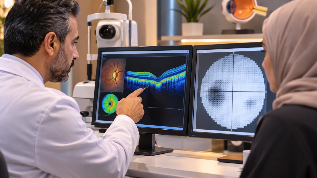
Your ophthalmologist may tell you that your eye pressure is normal but still recommend an optic nerve scan or a visual field test. This can raise an important question: Why are these tests necessary if your vision seems perfectly fine?
Glaucoma tests help detect changes in the optic nerve before noticeable vision loss occurs. Two of the most important diagnostic tools are Optical Coherence Tomography (OCT) and visual field testing. While OCT evaluates structural changes in the nerve fibers, visual field testing assesses how those changes may affect visual function.
Diagnosing glaucoma requires more than one examination. Ophthalmologists interpret several clinical findings together to determine whether glaucoma is present, assess the risk of developing the disease, and decide whether monitoring or treatment is necessary.
Why Are Glaucoma Tests Important for Early Detection?
Glaucoma refers to a group of eye conditions that progressively damage the optic nerve, which carries visual information from the eye to the brain.
The disease often develops without pain or noticeable symptoms during its early stages, particularly in open-angle glaucoma.
Early detection is essential because vision loss caused by glaucoma is generally irreversible. However, appropriate treatment can help slow disease progression and preserve remaining vision.
Importantly, elevated eye pressure does not always indicate glaucoma, and normal eye pressure does not necessarily rule it out. Understanding the difference between high eye pressure and glaucoma explains why ophthalmologists assess optic nerve health rather than relying on pressure measurements alone.

What Tests Are Used to Diagnose Glaucoma?
A comprehensive glaucoma evaluation may involve several examinations. The ophthalmologist selects the appropriate tests based on the patient’s clinical findings and individual risk factors.
1. Eye Pressure Measurement (Tonometry)
Tonometry measures intraocular pressure (IOP), one of the major risk factors associated with glaucoma.
An ophthalmologist may use a contact tonometer after applying anesthetic eye drops or another suitable measurement technique.
Goldmann applanation tonometry is widely regarded as a reference method for measuring intraocular pressure in clinical practice.
However, a single pressure reading cannot confirm or exclude glaucoma. Eye pressure fluctuates throughout the day, and individual optic nerves vary in their susceptibility to damage.
2. Optic Nerve Examination
During this examination, the ophthalmologist evaluates the optic nerve head for structural changes that may indicate glaucoma.
These may include thinning of the neuroretinal rim or increased cupping of the optic disc.
Optic disc photography may also be used to document the nerve’s appearance and compare findings during future appointments.
3. Optical Coherence Tomography (OCT)
OCT provides detailed measurements of the retinal nerve fiber layer and ganglion cell structures involved in transmitting visual signals.
These measurements help identify structural changes associated with glaucoma and monitor whether damage progresses over time.
4. Visual Field Testing (Perimetry)
Visual field testing measures the eye’s ability to detect visual stimuli across different areas of the field of vision.
It helps identify regions where visual sensitivity may have decreased because of optic nerve damage.
5. Gonioscopy and Corneal Thickness Measurement
Gonioscopy allows the ophthalmologist to examine the eye’s drainage angle and assess its anatomical characteristics.
Corneal thickness measurement, known as pachymetry, helps interpret intraocular pressure readings and evaluate glaucoma-related risk factors.
Together, these examinations provide a more comprehensive assessment than eye pressure measurement alone.
How Does OCT Detect Optic Nerve Damage?
Optical Coherence Tomography (OCT) is a non-invasive imaging technique that uses light to produce high-resolution cross-sectional images of the retina and structures associated with the optic nerve.
It allows ophthalmologists to examine subtle structural changes that may occur before patients notice a decline in vision.
OCT can help assess:
- Retinal Nerve Fiber Layer (RNFL) thickness: Measurements that may reveal thinning of nerve fibers associated with glaucoma.
- Macular ganglion cell layers: Evaluation of retinal cells involved in processing and transmitting visual information.
- Comparison with reference values: Identifying measurements that differ from expected ranges.
- Changes over time: Comparing previous and current scans to detect possible disease progression.
What Do OCT Results Mean in Glaucoma?
An OCT report may reveal thinning of the retinal nerve fiber layer or changes in optic nerve structure that suggest glaucomatous damage.
However, an abnormal OCT finding does not automatically mean a patient has glaucoma.
Results may be influenced by severe myopia, natural anatomical variations, imaging artifacts, or other eye conditions.
For this reason, ophthalmologists interpret OCT findings alongside clinical examination results, intraocular pressure measurements, and visual field testing.
The colors and numerical values displayed on an OCT report should never be interpreted in isolation.
OCT is also valuable for long-term monitoring, as repeated scans can help identify structural changes that may indicate progression.
How Is an OCT Eye Test Performed? Is It Painful?
An OCT scan is usually performed without touching the eye.
The patient places their chin and forehead against the device’s supports and looks at a fixed target while the machine captures detailed images.
The procedure is generally painless, and the imaging itself takes only a short time.
Occasionally, the technician may need to repeat a scan to obtain clearer images.
Depending on the examination, dilating eye drops may be used to improve the assessment.
How Does Visual Field Testing Detect Glaucoma-Related Vision Loss?
Unlike a standard visual acuity test, which measures how clearly you can read letters, a visual field test evaluates sensitivity across different areas of your vision.
This includes peripheral areas that you are not looking at directly.
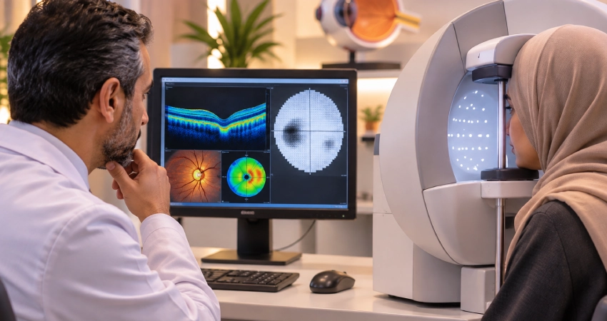
During the examination, the patient focuses on a fixed point inside the testing device and presses a button whenever a small light appears.
The test helps identify areas of reduced visual sensitivity or missing portions of the visual field.
These findings may indicate functional changes associated with optic nerve damage.
What Do Blind Spots on a Visual Field Test Mean?
Areas of reduced sensitivity or missing vision may suggest that the optic nerve is not functioning normally.
However, an abnormal visual field result does not confirm glaucoma on its own.
Fatigue, poor concentration, difficulty maintaining fixation, and other eye conditions can affect test accuracy.
An ophthalmologist may recommend repeating the examination, particularly when new findings do not match other clinical results.
Comparing visual field tests over time helps determine whether the condition remains stable or is progressing.
OCT vs. Visual Field Testing: What Is the Difference?
Although both tests are important for glaucoma evaluation, they assess different aspects of eye health.
| Feature | OCT | Visual Field Test |
| What it measures | Retinal nerve fiber and ganglion cell structure | Visual sensitivity across different areas |
| Type of assessment | Structural | Functional |
| How it works | Light-based retinal imaging | Responses to small visual stimuli |
| Role in early detection | May detect nerve fiber thinning | May identify unnoticed functional vision loss |
| Role in monitoring | Tracks structural changes over time | Tracks changes in visual field sensitivity |
Is OCT Better Than Visual Field Testing?
Neither test can fully replace the other.
In some patients, OCT may reveal structural damage before measurable visual field loss becomes apparent.
In other cases, visual field testing may detect functional abnormalities that are not clearly reflected in OCT measurements.
Combining structural and functional assessments provides a more complete picture of optic nerve health.
Can Glaucoma Be Detected When Eye Pressure Is Normal?
Yes. Some patients develop a condition known as normal-tension glaucoma, in which optic nerve damage occurs despite eye pressure readings remaining within the generally accepted normal range.
In these cases, optic nerve examination, OCT imaging, and visual field testing become particularly important.
Relying exclusively on eye pressure measurements may result in the disease being overlooked.
A diagnosis requires findings consistent with glaucoma and consideration of other possible causes of optic nerve damage.
For a more detailed explanation, our article on normal-tension glaucoma explores why optic nerve damage can develop even when eye pressure appears normal.
Who Should Have Glaucoma Testing?
Glaucoma examinations are particularly important for people with certain risk factors.
These include:
- Individuals with a family history of glaucoma.
- Older adults.
- People who have previously recorded elevated intraocular pressure.
- Patients with suspicious optic nerve findings.
- Individuals using corticosteroid medications for extended periods.
- People with severe myopia or additional glaucoma-related risk factors.
Having one or more of these risk factors does not necessarily mean glaucoma is present.
However, an ophthalmologist may recommend more frequent or specialized examinations depending on the patient’s individual circumstances.
A comprehensive eye examination at Batal Eye Center may involve more than checking visual acuity. Depending on clinical needs, it can include eye pressure measurement and evaluation of retinal and optic nerve health.
How Often Should OCT and Visual Field Tests Be Repeated?
Patients diagnosed with glaucoma, as well as those considered glaucoma suspects, may need repeated OCT scans and visual field tests.
The recommended frequency depends on factors such as:
- Glaucoma severity.
- Intraocular pressure levels and stability.
- Previous diagnostic findings.
- The rate of optic nerve damage, if progression has been identified.
Patients with unstable or progressing glaucoma may require more frequent testing.
Those with stable findings may be monitored at longer intervals, depending on their ophthalmologist’s recommendations.
Keeping previous OCT scans and visual field reports is important because comparing results over time often provides more meaningful information than interpreting a single examination.
Glaucoma Diagnosis and Optic Nerve Monitoring with Dr. Obaidah Kilani
Accurate glaucoma assessment requires experience in interpreting both structural and functional diagnostic findings, particularly when eye pressure is not elevated or optic nerve changes are still subtle.
Dr. Obaidah Kilani is a Consultant Ophthalmologist specializing in glaucoma diagnosis, treatment, and surgery, with more than 15 years of professional experience.
He is a Fellow of the Royal College of Ophthalmologists (FRCOphth), with expertise in advanced glaucoma procedures and minimally invasive glaucoma surgery (MIGS).
Specialized clinical experience supports the interpretation of diagnostic findings and helps determine whether a patient requires continued monitoring, initiation of treatment, or adjustments to an existing treatment plan.
Patients can learn more about his qualifications and professional experience through Dr. Obaidah Kilani’s profile on the official Batal Eye Center website.
Book a Glaucoma Examination at Batal Eye Center in Jeddah
Glaucoma may develop without noticeable vision changes, making specialized examinations essential for evaluating optic nerve health before significant visual problems occur.
At Batal Eye Center in Jeddah, patients have access to glaucoma diagnosis, monitoring, and treatment services.
The appropriate examinations are selected according to each patient’s medical history, risk factors, and clinical findings.
If you have a family history of glaucoma, previous examinations have identified suspicious optic nerve changes, or your ophthalmologist has recommended OCT or visual field testing, a specialized assessment may be appropriate.
Contact Batal Eye Center to schedule an appointment and discuss which glaucoma examinations are recommended for your condition.
Frequently Asked Questions About Glaucoma Tests
Can OCT Detect Glaucoma Early?
OCT may detect thinning of retinal nerve fibers associated with glaucoma before noticeable visual field loss develops in some patients.
However, OCT cannot confirm glaucoma on its own. Its findings must be interpreted alongside other clinical examinations.
Is Visual Field Testing Necessary for Glaucoma Diagnosis?
Visual field testing is an important part of glaucoma evaluation because it assesses how the disease affects visual function.
It also helps ophthalmologists detect changes and monitor disease progression.
Can OCT Results Be Normal Even If You Have Glaucoma?
Yes. Early glaucomatous changes may not always be clearly reflected in OCT measurements.
A normal scan does not completely exclude glaucoma when other clinical findings suggest the disease may be present.
Is an OCT Eye Scan Painful?
No. OCT is a non-invasive imaging procedure that generally does not require direct contact with the eye.
The scan is typically painless and takes only a short time.
How Long Does a Visual Field Test Take?
The duration depends on the type of test and the equipment used.
Common automated visual field tests usually take several minutes per eye, although additional time may be needed for breaks or repeated measurements.
Can Poor Concentration Affect Visual Field Test Results?
Yes. Fatigue, distraction, and difficulty maintaining fixation can affect test reliability.
The ophthalmologist reviews the reliability indicators and may recommend repeating the test if necessary.
Does Normal Eye Pressure Mean an Optic Nerve Examination Is Unnecessary?
No. Optic nerve damage can occur even when intraocular pressure falls within the normal range.
For this reason, pressure measurements alone cannot reliably exclude glaucoma.
Do OCT and Visual Field Tests Need to Be Performed at Every Appointment?
Not necessarily.
The ophthalmologist determines how often each examination should be repeated based on disease severity, stability, and previous results.
Can Vision Lost to Glaucoma Be Restored?
Vision loss caused by glaucoma is generally permanent because damaged optic nerve fibers cannot currently be restored.
However, early diagnosis and appropriate treatment can help slow further damage and protect remaining vision.