Ultrasound
Ultrasound
Ophthalmic ultrasound is one of the best and most accurate devices that we use at Batal Specialist Center to diagnose eye diseases and obtain a rapid assessment without surgical intervention. It helps our doctors to provide information and understand the correct treatment pathway and type of intervention.

Widefield sound wave device
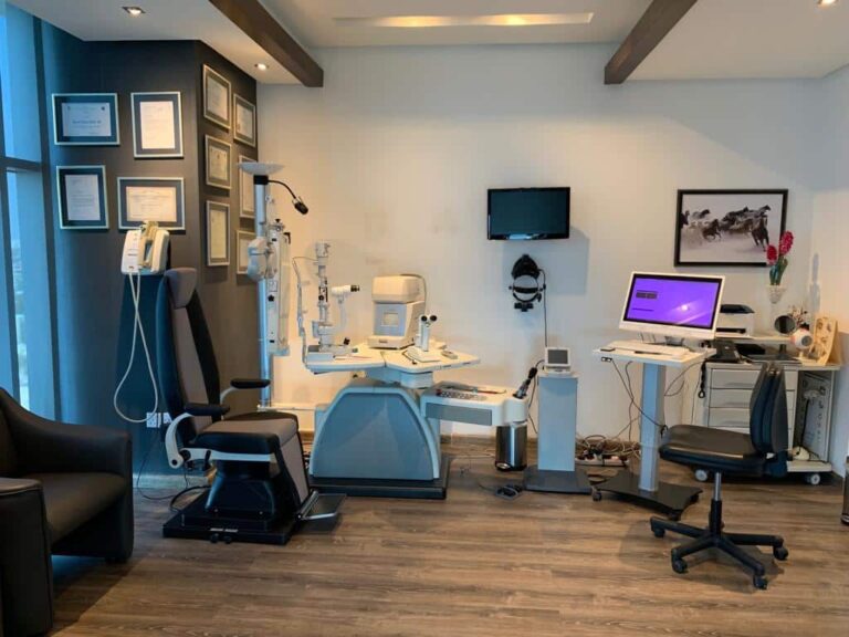
Batal Eye Specialist Center contains the latest ultrasound device with capabilities to examine the front of the eye and imaging the back of the eye to give details of the retina and vitreous.
When do we use the ultrasound device in Batal Center?
- An ultrasound is a simple and painless procedure that takes between 5 to 20 minutes. The eye does not need to remain open during the examination and it does not require any special preparations before it is performed, and children do not need anesthesia, so we use ultrasound in many cases.
What is ultrasound?
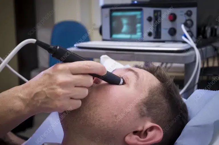
In most cases, not much needs to be done before the ultrasound examination. The patient lies on the examination table. A transparent watery gel is placed on the skin over the part to be examined (the eye in our case). This gel helps the sound waves pass through the body. Then a hand-held probe is moved. (“transducer”) above this part and there is no radiation hazard and the patient can return to daily tasks immediately after the test.
Sound waves to examine the eye
Sound waves to follow the surgery
Determine the type of eye bleeding
Diagnosing eye diseases
Usually, we only need light when a conventional eye examination, but the traditional examination may be difficult if there is blood in the eye or in cases of cataracts. In these cases, this type of ultrasound examination helps us to identify and diagnose eye diseases before determining the treatment, and it is used to discover:
Tumors or cysts of the eye.
Measuring the thickness and size of the tumor.
Retinal detachment.
Glaucoma Diagnosis.
eye lens darkening.
Follow-up after LASIK (lens implantation after removing the
natural lens to treat cataracts).
Foreign bodies in the eye, such as blood, lumps, or fatty bags on the iris.
Eye swelling.
Injury to the eye socket.
Detachment of the vitreous.
Eye length measurement.
Ultrasound features
Examination using high-frequency sound waves helps to examine soft tissues that are not visible on x-rays.
- The patient is not exposed to ionizing radiation and thus this examination is safer than other diagnostic techniques such as X-rays and CT scans.
- Less expensive than other methods
- Ultrasound is painless and does not require injections, punctures or anesthesia prior to the examination.
- The scan only takes several minutes.
- The scan results are immediate.
- Ultrasound imaging is done while the eyelid is closed, which facilitates the examination process in children or those with sensitive eyes.

Ultrasound Imaging | Doppler Device
Sound wave technology has advanced and provided various types of imaging, including Doppler, which is a special type of sound waves that measure blood flow through blood vessels in a fast and painless way to check for problems in the blood vessels and detect clots.
An accurate ultrasound test gives our doctors vital information to help them determine the best way to treat the eye. You can ask the doctor about whether the Doppler ultrasound affects the color of the eyes. In general, there are different types of Doppler scans, including:
Color Doppler: Determines the speed and direction of blood flow in real time / current.
Power Doppler: A new type of color Doppler and provides more details than the traditional color Doppler about blood flow, but it does not show the direction of blood flow, which may be important in diagnosing some cases, and here the spectral Doppler can be used
Spectral Doppler: It helps to show the percentage of blockage of blood vessels. It is not much different from a duplex doppler.
Doplex Doppler: Measures blood flow in a blood vessel and is not much different from Doppler spectroscopy.
Continuous Wave Doppler: allows more accurate measurement of blood flowing at higher speeds.
Doppler helps diagnose:
- Eye clots.
- Eye swelling.
- Weak blood vessels and thus fluid accumulation behind the eye.

How to book an ultrasound scan
You can book an ultrasound examination at the reception desk at Batal Specialist Center and you will not wait long before a specialist calls you, or you can make an appropriate appointment online.


.Your ophthalmologist will review the results with you and your doctor will make sure that the measurements of your eye taken from the scan are within the normal range.
A B test will give your doctor structural information about your eye. If results are abnormal, your doctor will need to determine the cause. Some conditions that a B test may reveal include:- Foreign bodies in the eye.
- Cysts.
- Swelling
- Retinal detachment
- Tissue damage or injury to the eye socket (orbit).
- Vitreous hemorrhage (bleeding into the clear gelatinous substance called the vitreous humor that fills the back of the eye).
- Cancer of the retina, under the retina, or other parts of the eye.
What is ultrasound of the eye?
This test provides a more detailed view of the inside of your eye than a routine eye exam. An ultrasound technician or ophthalmologist (a doctor who specializes in diagnosing and treating disorders and diseases of the eye) usually performs the procedure (sometimes called eye studies). Eye studies can be done in an office or imaging center. outpatient or hospital.
Why do I need an eye ultrasound?
This procedure is useful in identifying eye problems as well as diagnosing eye diseases. Some problems that the test can help identify include:
Tumors or tumors affecting the eye.
external influences.
retinal detachment.
Ultrasound of the eye and shell may also be used to help diagnose or monitor:
Glaucoma (a progressive disease that can lead to vision loss).
Cataracts (cloudy areas in the lens).
Lens implants (plastic lenses implanted in the eye after removal of the natural lens usually due to cataracts).
Do you need an A or B ultrasound?
A test: It uses high-frequency sound waves to measure the length of the eye, the curvature of the cornea, and determine the appropriate lens, for example, before cataract or cataract operation. It can be performed while sitting or while lying down.
B test: It helps to see and examine the area behind the eye. In some cases, cataracts and other diseases cause the back of the eye not to be seen by conventional screening devices, and here comes the role of ultrasound in diagnosing tumors, retinal detachment, and other conditions.

Senior Consultant, Pediatric Surgery and Eye Surgery Fellow of the British Royal Surgical College Fellow of the American Academy of Ophthalmology
Specialization: Strabismus and pediatric eye surgery
Degree: Member of the British Royal College of Surgeons and Member of the American Academy of Ophthalmology
Batal Eye Center
Call us 8001111897
Contact us via Email: in**@************er.com
Address: 8125 Prince Sultan road, Murjana tower Jeddah, Saudi Arabia


