

Med info
The Newest Vision Correction Techniques for Keratoconus

Keratoconus is a condition that affects the cornea, the clear, rounded front surface of the eye. Over time, the cornea gradually becomes thinner and weaker, eventually bulging outward into a cone-like shape. This abnormal change in shape distorts and blurs vision because it prevents incoming light from focusing properly on the retina.
It is estimated that keratoconus affects between 1 in 500 to 1 in 2,000 people worldwide, although it may be more common in certain populations. This article from Batal Specialized Medical Complex explains modern vision correction options for keratoconus and the available treatments.
What Is Keratoconus?
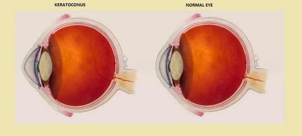
Keratoconus is an eye condition that affects the cornea, the transparent front part of the eye. In a healthy eye, the cornea has a smooth, dome-like shape. With keratoconus, the cornea gradually thins and begins to protrude forward into a cone shape.
This changes how the eye focuses light and leads to distorted, blurred vision.
Keratoconus usually appears in younger people between the ages of 16 and 30 and may continue to progress until around the age of 40. It typically affects both eyes, although one eye is often more severely affected than the other. If you notice any eye symptoms or changes in your vision, you should see an ophthalmologist.
Symptoms of Keratoconus
If you have keratoconus, you may experience certain symptoms. In this section, we will outline the most common signs and symptoms of keratoconus.
How a Keratoconus Patient Sees
The exact cause of keratoconus in some individuals is still unclear, but we know that symptoms arise from the distorted shape of the cornea and from scarring that can occur at the highest points of the cone.
Early symptoms often present as simple blurred vision, similar to common refractive errors like myopia or regular astigmatism.
However, the hallmark of keratoconus is “ghosting” or multiple shadow images around objects. This may also appear as glare, halos, or starbursts around lights.
Patients frequently complain of light sensitivity (photophobia), occasional itching, and generally poor night vision. Visual distortion and blur are usually more noticeable in dim lighting, because the pupil dilates to let in more light and exposes a larger, more irregular corneal surface.
When Is Vision Correction for Keratoconus Performed?
Keratoconus typically develops sometime between the early teenage years and the early twenties and gradually worsens over time. If you are diagnosed with keratoconus, you may wonder how it affects your eligibility for LASIK or other laser eye surgeries.
Keratoconus is a progressive thinning and bulging of the cornea, the eye’s clear front “window.” A normal cornea is round and regular, while a keratoconic cornea bulges outward like a cone.
Vision can be affected in two main ways:
– The formerly smooth corneal surface becomes irregular, causing irregular astigmatism and/or increasing myopia.
– Symptoms can include double vision, glare, halos or starbursts around lights, and difficulty seeing in certain lighting conditions.
If you have keratoconus (even in its early or mild stages), most laser eye surgeons will advise against laser vision correction.
Procedures such as LASIK and PRK reshape the cornea by removing microscopic amounts of corneal tissue. Removing tissue from an already thin and structurally weak cornea further destabilizes it, compromises corneal integrity, and can severely worsen vision. For this reason, your ophthalmologist will usually recommend eyeglasses or contact lenses to correct your vision instead.
Unfortunately, keratoconus can make it difficult to wear contact lenses comfortably or for extended periods, especially with standard soft lenses. Another common warning sign is the need for frequent changes in your glasses or contact lens prescription, requiring multiple visits for adjustments. Understandably, many patients begin to look for a more permanent solution.
The Latest Advances in Keratoconus Treatment
One of the most important advances in the management of keratoconus is the development of intrastromal corneal ring segments (ICRS), commonly known by a brand name such as Intacs™.
Intacs are tiny, semi-circular inserts implanted within the cornea to help reshape its curvature. The primary goal of Intacs is not to eliminate your need for glasses or contact lenses, but rather to:
– Regularize the corneal shape,
– Improve the quality of vision,
– And allow you to wear contact lenses more comfortably and for longer periods.
Although there is no absolute “cure” for keratoconus, disease progression can often be halted with a procedure known as corneal collagen cross-linking (CXL). A cornea specialist can determine whether CXL and/or Intacs are suitable for your specific case.
If you are experiencing the effects of keratoconus and would like to discuss Intacs or other advanced treatments, please contact the specialized eye center at Batal Medical Complex in Jeddah.
Diagnosing Keratoconus at Batal Medical Complex in Jeddah
To diagnose keratoconus, your doctor needs to accurately measure the shape and thickness of your cornea. There are several methods to do this, but the most common is called corneal topography.
During corneal topography, the doctor takes detailed images of your cornea and analyzes the curvature patterns to detect even very early or subtle changes.
Children of parents with keratoconus should have their eyes examined once a year starting around age 10, since early detection allows for earlier intervention.
Treatment often begins with a new glasses prescription. In mild cases, glasses may provide satisfactory vision. If they do not, your doctor will likely recommend contact lenses. Rigid gas permeable (RGP) or other types of specialty hard lenses are usually the first choice. Over time, you may need additional treatments to strengthen the cornea and improve your vision, including advanced keratoconus vision correction procedures.
Advantages of Vision Correction for Keratoconus
Keratoconus typically begins in adolescence or early adulthood, and the corneal changes slowly worsen over time. Symptoms tend to stabilize once the corneal shape stops changing, which usually happens roughly 20 years after onset.
(You may also be interested in: Femto-LASIK Vision Correction Surgery)
Vision Correction Procedures for Keratoconus
In a healthy eye, the cornea has a smooth, dome-like shape. In a patient with keratoconus, the cornea becomes thin and weak and bulges outward into a cone. This can occur in one eye or both and often begins in the teenage years or early twenties. You may experience blurred or distorted vision, and your nearsightedness may progressively increase.
In some people, keratoconus is only discovered during a preoperative evaluation for laser refractive surgery, because it may otherwise cause few or no obvious symptoms in the early stages. Although this means you are not a candidate for standard laser eye surgery, there are other treatment options to help preserve and support your vision over the long term.
The treatment approach depends on the stage and stability of your keratoconus:
– If your keratoconus is stable, implantable collamer lenses (ICLs) may be used to improve your vision.
– We do not recommend laser eye surgery (such as LASIK) as a refractive procedure for patients with keratoconus, because it is a degenerative disease that thins the cornea.
– Laser refractive procedures like LASIK remove tissue from an already thin, weakened cornea, which can cause further thinning, unpredictable changes in corneal shape, and potential loss of vision.
In selected cases, a combined approach known as “Refractive CXL” or “CXL-plus” may be used. This combines PRK (a surface laser procedure) with corneal cross-linking to:
– Improve the quality of vision,
– Stabilize the cornea.
Even after this combined procedure, you will usually still need glasses or contact lenses for best vision. However, bothersome night-vision problems such as halos, glare, and starbursts often improve.
For carefully selected patients whose condition is stable after CXL-plus, implantable contact lens (ICL) surgery may be offered to reduce or eliminate dependence on glasses or contact lenses.
Once the cornea has been stabilized, additional options such as laser vision correction (in very specific, cautious scenarios) or refractive lens exchange may be considered to further reduce dependence on visual aids.
Comprehensive eye examinations at our center allow us to detect keratoconus and its earliest changes, even before significant symptoms develop.
(You may also be interested in: Corneal Astigmatism and Vision Correction at Batal Complex in Jeddah)

Vision Correction for Keratoconus
LASIK is a popular refractive surgery that has helped millions of people achieve clear vision without glasses or contact lenses. However, if you have been diagnosed with keratoconus, it is natural to ask whether LASIK is still an option.
Keratoconus affects the shape of the cornea, causing it to become thin and bulge outward into a cone. This leads to blurred, distorted vision and can be quite uncomfortable.
Unfortunately, LASIK is not a safe or appropriate choice for patients with keratoconus. The procedure works by reshaping the cornea, removing a thin layer of corneal tissue with a laser. In keratoconus, the cornea is already thin and structurally weak. Removing additional tissue can further destabilize the cornea, worsen the disease, and potentially lead to serious vision problems.
If you have keratoconus, standard LASIK is generally not an option. However, there is another surgical approach that can offer effective vision correction in selected cases.
Implantable collamer lens (ICL) surgery involves placing a small, biocompatible lens inside the eye, between the iris and the eye’s natural lens, to improve vision. The ICL is custom-designed to match your eye’s shape and prescription. Once in place, it works together with your natural lens to provide clear vision, without reshaping the cornea.
ICL surgery is considered a safe and effective option for many patients with mild keratoconus, because it does not involve removing corneal tissue. However, in most modern treatment protocols, you must first undergo corneal cross-linking to stabilize the keratoconus before you can be considered for ICL implantation.
(You may also be interested in: Benefits of Laser Vision Correction – A New Clarity for Better Sight)

How Long Does Keratoconus Vision Correction Take?

The procedure commonly referred to as “contact lens surgery” in this context is corneal cross-linking (CXL), often combined with a temporary bandage contact lens. The treatment itself typically takes about 60–90 minutes.
A bandage contact lens is placed on the eye at the end of the procedure to protect the cornea and aid healing. This lens is usually left in place for about a week. After the procedure, patients often experience burning, irritation, or discomfort in the eye. Mild to moderate discomfort is expected, but if the pain is severe, you should contact your doctor immediately.
Success Rate of Keratoconus Vision Correction
Approximately 19 out of 20 corneal transplants performed for keratoconus are successful and remain clear for at least five years. If you have advanced keratoconus and can no longer tolerate or achieve good vision with contact lenses, corneal transplantation is often the best treatment option.
Overall, vision correction approaches for keratoconus—whether cross-linking, ring segments (Intacs), ICLs, or, in advanced cases, corneal transplant—offer excellent outcomes. Reported success rates for appropriately selected keratoconus vision correction procedures can exceed 96%.
Postoperative Follow-Up at Batal Specialized Complex
The treated eye is usually sore or uncomfortable for about 3 to 5 days after the procedure, although the level of discomfort varies from one patient to another. Initial healing typically takes around one week, though some patients may feel that full visual recovery takes a bit longer.
Regardless of the type or degree of your visual problem, keratoconus and other corneal conditions require careful evaluation and a tailored treatment plan from an experienced ophthalmologist, using effective, up-to-date medical tools and technology.
Whatever eye condition you may be facing, you can book an appointment at Batal Specialized Complex for a thorough eye examination and to begin addressing your vision problems, no matter how complex they may seem.
Frequently Asked Questions About Vision Correction for Keratoconus
Does vision correction work in keratoconus?
If you have keratoconus (even mild keratoconus), laser eye surgeons will generally discourage you from having laser vision correction. Procedures such as LASIK and PRK reshape the cornea by removing microscopic amounts of corneal tissue, which can further weaken an already thin cornea.
Can I correct my vision after corneal cross-linking?
Vision correction after corneal cross-linking requires a detailed, in-person evaluation. Your ophthalmologist must carefully assess corneal stability, thickness, and shape before deciding whether additional procedures such as ICL implantation or other options are appropriate.
Is keratoconus a serious disease?
We see through the cornea, which is the clear central part of the eye’s front surface. Normally, the cornea has a smooth, round, dome-like shape. In some people, however, the corneal structure is not strong enough to maintain this shape. Over time, the normal curve of the corneal surface bulges outward into a cone. This condition is called keratoconus.
Keratoconus is usually detected in the teenage years or twenties but can also begin in childhood. In some cases, it is only diagnosed later in life, often when it is mild. Changes in corneal shape occur over several years, but they tend to progress faster in younger patients.
If left untreated, keratoconus can lead to permanent loss of vision. The corneal irregularities make it hard for the eye to focus with or without glasses or standard soft contact lenses.
Does phone use worsen keratoconus?
There is no strong evidence that using computers or digital devices such as tablets, e-readers, or smartphones directly worsens keratoconus. However, excessive use of digital screens can contribute to dry eye symptoms, which may add to general eye discomfort.
At what age does keratoconus usually stop progressing?
Keratoconus can appear between the ages of about 10 and 25 and usually progresses slowly until around age 40. In many patients, the disease tends to stabilize around this age. Even after progression slows or stops, treatment options are available to help manage symptoms and improve vision.
When does keratoconus stop progressing?
Keratoconus involves thinning and steepening of the cornea, the part of the eye that helps focus incoming light onto the retina. As the cornea gradually becomes more cone-shaped, vision can become severely blurred and other visual symptoms may appear.
The disease can progress quickly in some cases or slowly over many years. Early detection and timely intervention can slow or halt progression and reduce long-term impact.
Another option to improve vision and partially reverse corneal irregularity is the use of small implants called Intacs. These are inserted into the cornea to reshape its curvature from within. By flattening the steepest part of the cone, Intacs can significantly reduce some of the uncomfortable visual symptoms of keratoconus.

The exact cause of keratoconus is still not fully understood. Most specialists believe there is a strong genetic component. It often begins to develop during adolescence and tends to stop progressing by the age of about 30 to 40 in many patients. Nonetheless, monitoring and appropriate treatment remain important throughout the course of the disease.


