

Med info
The Cornea in Detail: Structure, Function, and What Affects It
The cornea is a crucial part of the eye that helps you see clearly. If you want to understand how your eyes work, how vision happens, and you’re looking for information about the cornea, it’s helpful to know what the cornea is, how it functions, and what can harm it. Keep reading to learn more about the cornea and how you can protect your eye health.
Also read: When Does Vision Stop Getting Worse – Is That True?
What Is the Cornea?
The cornea is the clear, front surface of the eye. Because it’s transparent, light can pass through it. It covers the pupil, the iris, and the anterior chamber, and it’s made up of cells and proteins, not blood vessels.
The cornea gets its nutrients from the tear film and oils that spread across the surface every time you blink. This tear film layer protects the eye from debris and germs.
The cornea acts as a barrier that traps dust, dirt, and pollen between your eyelid and the corneal surface. Then your tears wash this debris off to the side where it’s blinked away, or you may see small particles and remove them manually.
Also read: Nearsightedness (Myopia) – Everything You Need to Know
Corneal Structure and Layers
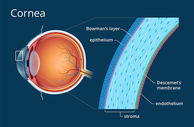
The cornea is made up of five main layers:
Epithelium
The epithelium is the outermost layer of the cornea. This smooth surface absorbs oxygen and contains thousands of nerve endings.
Bowman’s Layer
Bowman’s layer is the second layer of the cornea. It consists of strong, flexible collagen fibers within a clear tissue. When this layer is injured and then heals, it forms scars, and those scars can affect your vision.
Stroma
The stroma makes up about 90% of the corneal thickness. It is composed mainly of water and collagen.
Descemet’s Membrane
Descemet’s membrane is a thin layer that helps prevent infections and injuries from penetrating deeper into the eye and affecting the iris. It’s made of collagen and endothelial cells and has the ability to regenerate if your eye is injured.
Endothelium
The endothelium is the innermost layer of the cornea. It consists of a thin sheet of endothelial cells that keep the cornea clear by pumping excess fluid out of the stroma.
The endothelium is essential and does not regenerate if it’s damaged by trauma or disease. However, this layer can be replaced with a partial-thickness corneal transplant.
Also read: Farsightedness – What It Is, Causes, and Treatment
How Does the Cornea Work?
About 65–75% of the eye’s focusing power comes from the cornea. It works like a camera lens, allowing light to enter the eye. Light hits the cornea and is bent (refracted) as it passes through to the lens.
The lens then fine-tunes the focus of the light and directs it onto the retina. The retina converts light into electrical signals that travel to the brain through the optic nerve. The brain then interprets these signals as images you can understand and recognize.
Also read: Vision Screening Tests – What They Are and Types
What Conditions Can Affect the Cornea?
Also read: Low Vision – Symptoms and Everything You Need to Know
Keratoconus
Keratoconus is a corneal disorder that changes the shape of the cornea. A healthy cornea is normally smooth and slightly rounded. In people with keratoconus, the cornea becomes thinner, bulges forward, and takes on a cone-like shape.
When the cornea loses its smooth, regular curve, it thins out and becomes structurally weaker. This makes you more prone to disease and injury and can lead to blurred or distorted vision and light sensitivity.
There are several treatment options for keratoconus, depending on how advanced the condition is. In early stages, eye doctors often start with rigid gas permeable (RGP) or other specialty hard contact lenses.
These rigid lenses help maintain a healthy layer of tears on the eye, continuously bathing the cornea in moisture while you wear them. They protect the corneal surface from irritation, severe dryness, and minor trauma. Another treatment your eye doctor may consider is corneal cross-linking.
Corneal cross-linking is a procedure that helps stop keratoconus from getting worse by strengthening the cornea. It uses ultraviolet (UV) light and riboflavin (vitamin B2) eye drops to reinforce the collagen fibers in the cornea. For patients with advanced keratoconus, a corneal transplant may be needed to preserve their remaining vision.
Also read: Corneal Inflammation – Symptoms of Keratitis and Key Information
Fuchs’ Dystrophy
Fuchs’ endothelial dystrophy (often called Fuchs’ dystrophy) affects about 2% of people over age 60 and is the most common type of corneal dystrophy in many countries. This disease gradually damages the innermost layer of the cornea, the endothelium, leading to corneal swelling and blurred vision.
Severe cases require advanced forms of corneal transplantation to replace this thin layer of tissue. These procedures are called DMEK (Descemet Membrane Endothelial Keratoplasty) and DSAEK (Descemet Stripping Automated Endothelial Keratoplasty). Instead of a full-thickness corneal transplant, these advanced techniques selectively replace only the inner endothelial layer with donor endothelial tissue.
Also read: Requirements for Vision Correction Surgery – Are You a Candidate?
Corneal Ulcer
Corneal ulcers, a form of infectious keratitis, occur more often in people who wear contact lenses or have severe dry eye. These infections develop when bacteria, fungi, or viruses invade and damage the cornea.
Treatment for corneal ulcers usually includes antibiotic, antifungal, or antiviral eye drops, depending on the cause. You may also need pain relievers. Without prompt treatment, corneal ulcers can lead to permanent vision loss.
Also read: Specialized Eye Clinics – Top Ophthalmologists in Jeddah
Dry Eye Syndrome
Dry eye syndrome describes a condition in which patients either don’t produce enough tears, or their tears are of poor quality. When your eyes feel dry, it’s often because there isn’t enough water or oil in the tear film.
The tear film has three main components: water, oil (lipid), and mucus. If any one of these is lacking, the tears will not adequately lubricate and protect the surface of the eye. Dry eye syndrome is increasingly common in people who spend long periods looking at screens.
When you use digital devices like computers and smartphones, your blink rate drops, which reduces how often fresh tears spread over the eye and lowers overall lubrication.
You may also develop dry eye as a side effect of certain medications, from blockage of the meibomian glands (oil glands in the eyelids), or from a natural decline in tear production with age. When the eye doesn’t have enough tears, the cornea becomes more vulnerable to irritants such as dust and foreign particles. The corneal surface can then develop erosions, ulcers, and infections due to dryness, and if left untreated, this can result in vision loss.
Corneal Erosion or Recurrent Corneal Erosion Syndrome
Scratches on the cornea (corneal abrasions) can cause corneal erosion. Recurrent corneal erosion can happen spontaneously after an initial injury. If you previously scratched your eye—for example by rubbing it too hard—you may be more likely to experience future erosions at the same weak spot.
These erosions can cause blurred vision, light sensitivity, tearing, and redness in the eyes.
Also read: Keratoconus Surgery

Whatever type of visual impairment, refractive error, or eye problem you have, these delicate medical conditions require a thorough examination and appropriate treatment from a skilled ophthalmologist using effective medical tools and technology that deliver real results. Whatever eye disease you’re dealing with, you can book an appointment at Batal Specialized Medical Complex for a comprehensive eye exam and to begin managing and treating your eye problems, no matter how complex they are.


