

Med info
Retina: anatomy, function, diseases, and treatment methods
Vision is important in our lives because it connects us to our surroundings and allows us to stay safe, learn, and create memories. The retina is an important component of the eye because it transmits light entering the eye to the optic nerve, which in turn sends information to the brain. Any damage to the retina can lead to poor vision. Book with an ophthalmologist at Batal Specialist Complex immediately if you notice any changes in your vision.
The retina is the deepest layer of the eye. It consists of photoreceptor cells that convert light energy into nerve impulses. These electrical signals are passed through the optic nerve to the visual cortex, allowing us to visualize our surroundings.
In this article, we will focus on exploring the retina, its function, and its components. We will also discuss the consequences of retinal disease.
What is the retina and where is it located?
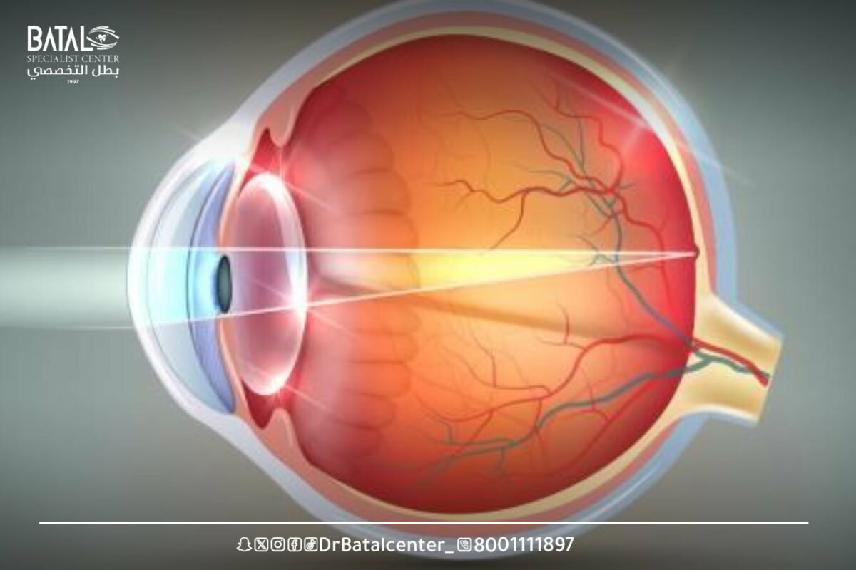
The retina is a thin layer of tissue that lines the back wall of the eye. This layer of tissue detects light and transmits messages to the brain, allowing you to see.
When light passes through the lens at the front of the eye and hits the retina, photoreceptors (light-sensitive cells in the retina) convert light energy into an electrical signal carried by the optic nerve and transmitted to the brain.
Thus, the neurons in the retina are responsible for all forms of vision, allowing you to see in low-light conditions, perceive the full range of colors and sharp edges of sensitive images, and have a wide field of view.
Retina in English
The medical term for retina in English is Retina.
Where is the retina located?
The retina is located at the back of the eye, forming the deepest border of the other layers of the eye.
The function of the retina and its role in vision
The retina contains millions of cells that work together to detect light, convert it into electrical signals, and communicate with the brain to produce vision. These small photoreceptor cells are called cones and rods. Together, cones and sticks give you the ability to distinguish between light and dark, as well as colors.
The function of the retina in the eye is to convert the light received by the eye into electrical signals. These signals then travel to the brain via the optic nerve, where it forms images. The main function of the retina is to help a person see.
What is a healthy retina made of?
The retina usually appears red or orange because it is surrounded by many blood vessels and consists of the following components:
Macula: The macula is a small but vital part of the central retina, and the macula is essential for seeing details of objects in front of you, such as faces and written text.
Click: The depression in the center of the spot, also known as the central click, is the sharpest point in the center of your vision. The fovea is perhaps the most important part of the human eye.
Peripheral retina: Peripheral retina is the retinal tissue outside the macula. It provides us with peripheral (side) vision and visibility in dim lighting settings.
Photoreceptors: Photoreceptors are located in the outer layer of the retina. Photoreceptor cells are neurons that convert light into an electrochemical signal, which is the first and crucial step in the process of perceiving light and color. Photoreceptor cells are divided into two types: rods and cones.
Sticks: These photoreceptor cells are very sensitive to low light levels and contribute to our night vision. They are located in peripheral retinal areas and are therefore also essential for peripheral vision.
Cones: Cone cells are photoreceptors that sense and process red, blue, and green colors to provide full-color vision. Cones are responsible for detailed vision of bright light and color vision.
Common retinal diseases and their causes
There are many retinal diseases that can affect your vision, some of which are described below:
Diabetic retinopathy
This common diabetic retinopathy occurs when high blood sugar levels damage the blood vessels in the retina. When blood vessels in the retina are damaged, they can leak, bleed, or close, all of which can lead to vision loss. Sometimes, pain may occur if blood access to the eye becomes more compromised. It is very important that every diabetic patient is examined at least once a year by an ophthalmologist at the Batal Specialist Complex.
Age-related macular degeneration
Age-related macular degeneration AMD: It is the leading cause of vision loss in adults over 50 years of age. Individuals with AMD experience loss of central vision and difficulty reading or seeing objects in front of them. AMD almost never causes complete blindness, but it can significantly impact daily life.
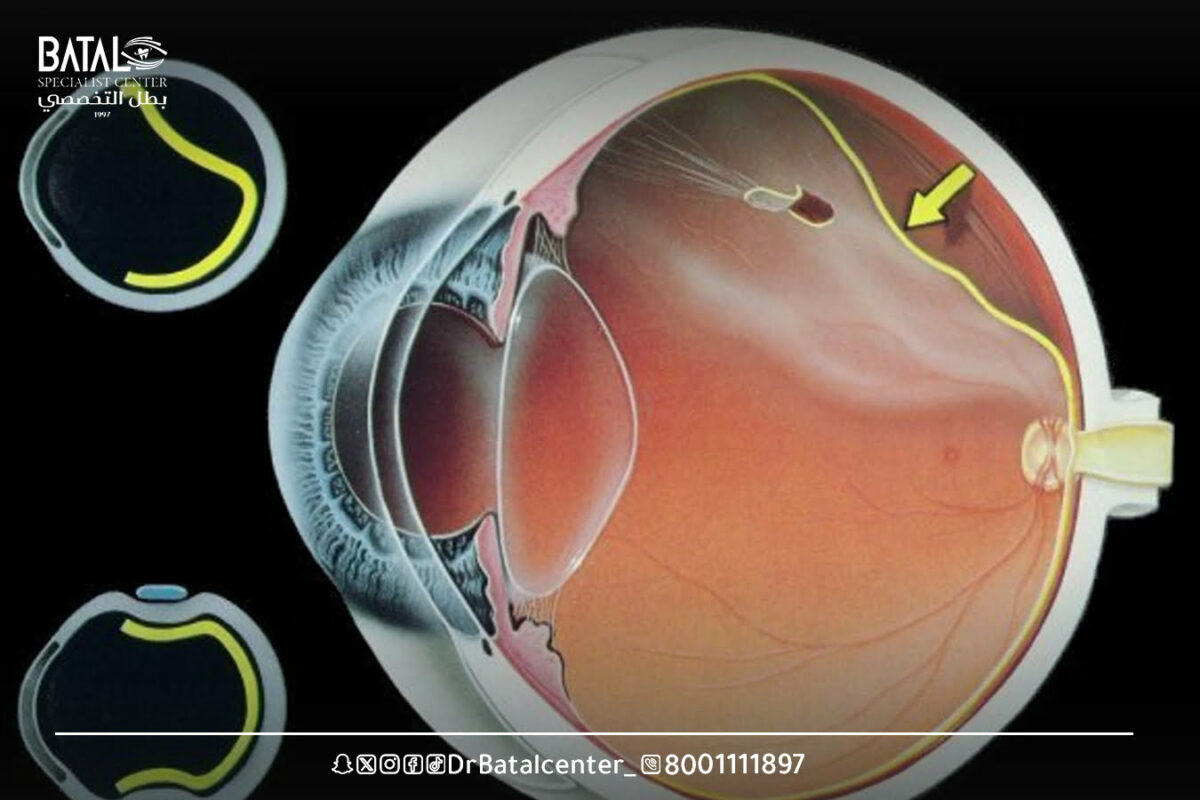
Retinal detachment
Retinal detachment is a serious eye disorder that impairs vision and, if left untreated, often leads to blindness. This occurs when retinal tissue separates from the underlying eye wall to which it normally attaches. This condition is often spread as a result of genetic factors, but can also result from trauma (for example, a blow to the head) or eye surgery. Retinal detachment is painless, but warning signs include new floaters, flashes of light, or loss of peripheral vision.
Macular hole
A macular hole is a small hole in the spot. The piercing may occur on its own or develop due to an eye injury.A macular hole usually causes vision problems in the center of vision and usually requires surgery to repair it.
Retinitis pigmentosa
Retinitis pigmentosa (RP)– is a genetic disorder that changes the retina’s sensitivity to light, making vision difficult. Depending on a person’s condition, the type and rate of vision loss associated with RP varies from person to person.
Supraretinal membrane
With age, when vitreous gel separates from the retina, it can sometimes leave some gel cells on the surface of the macula (center of the retina). These cells can multiply to form a membrane of scar tissue (macular wrinkle), which can distort and wrinkle the macula, resulting in blurry, distorted vision. If the scar membrane is large, surgical removal may be required in the operating room.
Tumors
Retinal carcinomas occur when nerve cells in the retina transform, multiply, and form a tumor. Cells usually spread throughout and around the eye, and can spread to other areas of the body, including the brain and spine. Retinoblastoma most often affects young children; however, it can sometimes affect adults.
Retinal treatment options at Batal Specialized Complex
The primary goal of treatment is to stop or delay the progression of the disease and maintain, improve, or restore vision. Sometimes the damage already done is irreparable, making early detection crucial. The Batal Specialist Complex physician will work with you to determine the best course of action. Treatment for retinal disease can be complex and, sometimes, urgent. Treatment options may include:
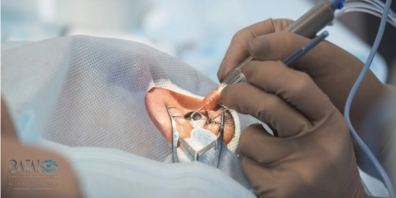
Surgery and vitrectomy
The best treatment for retinal detachment usually requires surgical repair. This can be achieved through a technique called vitrectomy, where the surgeon removes the gel that fills your eye and replaces it with oil or gas to push the retina back into the eye wall. Another technique involves pushing the eye wall inward with silicone tape to encourage retinal reattachment.
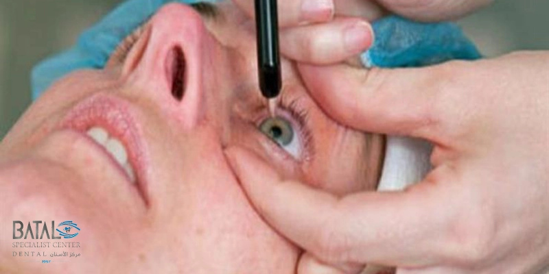
Medications and intraocular injections
Injecting the medication into the eye – Your doctor may suggest injecting the medication into your eye. This technique may be useful in treating patients with wet macular degeneration, diabetic retinopathy, and retinal vein occlusion.
Laser treatment
Retinal tear can be repaired using laser surgery. The surgeon will heat small points on the retina, which helps connect the retina to underlying tissue.
Prompt laser treatment of a new retinal tear can reduce the likelihood of it developing into retinal detachment.
Laser surgery can also be helpful in shrinking abnormal blood vessels in the bleeding retina, a technique called total retinal photocoagulation. Patients with diabetic retinopathy may benefit from this treatment.
The role of cone cells in the retina
Cones are found in photocyte receptors, or the central part of the retina. These cells enable you to see colors and help you see fine details. There are three types of cone cells: red, green, and blue. They work best in bright light and are responsible for high-definition vision that allows you to read and drive.
The best ways to prevent and strengthen the retina
The different ways in which a person can care for the retina mostly revolve around taking care of the overall health of the eye and the general body. Methods of caring for the retina and maintaining eye health include conducting eye examinations with an ophthalmologist at Batal Specialized Complex in Jeddah, eating a healthy and balanced diet, exercising regularly, and quitting smoking. Here are the ways to care for the retina:
- Attending eye tests with an ophthalmologist: Attending eye tests with an ophthalmologist is an important step in macular care and overall eye health. During an eye test, an ophthalmologist evaluates a person’s vision and examines the retina, including the retina. If there is a problem with the retina, an ophthalmologist will be able to detect it and provide appropriate treatment.
- Eat a healthy, balanced diet: Eating a healthy, balanced diet is an important step to help take care of eye structures, including the retina, and a person’s overall health. Eat a healthy and balanced diet that includes eating leafy vegetables, omega-3, fruits and vegetables, which helps provide the body with important vitamins and minerals, such as vitamin A and vitamin C. These vitamins can contribute to maintaining good eye health.
- Exercise regularly: Exercising regularly is an important measure to take to take care of overall health, which can contribute to good eye health. Regular exercise involves participating in activities such as going to the gym, exercising, or running, which helps maintain weight and reduces the chances of developing certain diseases.
- Quit smoking: Quitting smoking is an important way to take care of your overall health, as well as your eyes and retina, as smoking can increase your risk of age-related macular degeneration.
Regular retinal examinations at Batal Specialized Complex
Most children and adults should undergo a comprehensive eye examination every one to two years at the Batal Specialist Complex. Individuals at higher risk for eye disease may need to have their eyes checked more frequently. You may need to have more eye exams more frequently if you:
- They are over 60 years old.
- You have a family history of eye disease.
- Wear thick glasses.
- You have a health condition that can cause eye problems, such as diabetes.
- You have had eye surgery or suffered eye damage as a result of a stroke.
- During a comprehensive eye exam, your eye doctor will examine your eyes and perform several tests. Some of these tests help evaluate your vision and determine if you need glasses or contact lenses.
Treating retinal damage
Treatment for retinal damage can include various methods, including retinal transplantation and gene therapy, as follows:
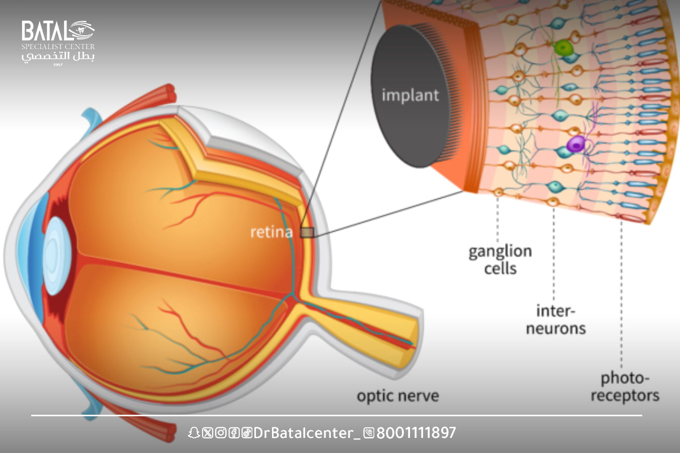
Retinal implant
Retinal transplantation involves the surgical replacement of damaged retinal tissue with healthy donor tissue to restore visual function. There are two main types of retinal implant techniques:
Autotransplantation: In this approach, healthy retinal tissue is taken from a different area of the patient’s retina and transplanted into the affected area. This technique is limited to the availability of donated healthy tissue inside the patient’s eye.
Allogeneic transplantation: Allogeneic transplantation involves the use of donor tissue from a different individual. Donor tissue is usually obtained from deceased donors or from embryonic stem cells. This technique provides an unlimited source of donor tissue, but comes with challenges related to immune rejection and integration of transplanted cells into the recipient’s retina.
Gene therapy
Gene therapy aims to treat retinal diseases by delivering therapeutic genes to the retina to correct genetic defects or provide therapeutic effects. There are different methods of gene therapy for retinal diseases:
Gene replacement therapy: This approach is used to treat genetic conditions in which a particular gene is defective or missing. The therapeutic gene is introduced into the retina using a viral vector, such as adeno-associated virus (AAV). The viral vector delivers the functional gene to retinal cells, allowing them to produce the lost protein and restore normal function.
Gene editing: Gene editing techniques are being explored to correct specific genetic mutations directly in a patient’s retina. This approach holds great potential for treating inherited retinal diseases caused by single genetic mutations.
Restore your visual health with retinologists at Batal Specialized Complex
At Batal Specialized Eye Complex, we care for the health of your retina using the latest diagnostic and treatment techniques under the supervision of doctors specializing in retinal and optic nerve diseases. Whether you suffer from diabetic retinopathy, retinal detachment, or poor vision, we provide you with comprehensive care that ensures the best visual outcomes.
Don’t wait for your vision to be affected — Book your appointment today for a comprehensive retinal examination at Batal Specialized Complex in Jeddah and prepare for clear vision with confidence.


