

Med info
Optic Nerve Examination
A visual acuity test evaluates several aspects of eye function. It gives an indication of the visual integrity of the eyes, the health of the retina, and the brain’s ability to interpret images.
This test is usually performed in a well‑lit environment, with the patient standing or sitting at least 6 meters (20 feet) from a Snellen chart (a board displaying several rows of letters that progressively decrease in size from top to bottom). If the patient normally wears distance glasses, they should wear them for this test. One eye is covered, and the patient is asked to read the letters in each row from top to bottom until they can no longer correctly identify the characters. The same process is then repeated for the other eye.
Each line on the chart is assigned a number that represents the distance at which a person with normal vision should be able to read letters of that size. For example, the largest letter at the top of the chart can normally be seen clearly at 60 meters. The distance between the patient and the chart (in this case, 6 m) is written as the numerator, and the number assigned to the smallest line the patient can read is written as the denominator.
If the patient is unable to read the letters on the 6‑meter line, a pinhole occluder should be placed in front of their glasses to correct for refractive error. For patients who cannot see the top letter at 6 meters, the chart should be moved to a distance of 1 meter. If this is still not helpful, the examiner assesses whether the patient can count fingers only, detect hand movements, or has only light perception.
In such cases, and also for children, alternatives to the Snellen chart can be used, such as charts with different shapes and sizes instead of letters.
See also: Benefits of Cold Compresses for the Eyes
Color Vision Testing
Color vision is best assessed using Ishihara plates. This test evaluates the patient’s ability to distinguish red and green colors. The examiner presents the plates to the patient and asks them to identify the numbers on each plate, which are designed as mosaic‑like patterns of different shades of red and green.
The first plate in the set is a screening plate that tests visual acuity. If the patient is unable to identify the number on this first plate, the problem is likely with visual acuity rather than color perception. A more basic way to assess red‑green color blindness is to present a solid block of red or green color (these are sometimes printed at the bottom of some Snellen charts).
Red desaturation is one of the earliest signs of optic nerve involvement and indicates reduced ability to distinguish red objects. Other red‑green color vision problems may be congenital and are typically X‑linked disorders that occur more commonly in males. Acquired red‑green desaturation can extend from the photoreceptor layer in the retina to the lateral geniculate nucleus in the thalamus.
Visual Field Testing
The interpretation of a visual field test depends in part on the examiner’s own visual fields, as some aspects of the test are based on comparison with the examiner. There are several important features of the visual fields that should be assessed:
See also: Causes of Blurred Vision and Everything Related to It
Lesions and Defects
Some of these defects include the following:
You are advised to visit the Eye Center at Al Batal Specialized Hospital in Jeddah, which is distinguished by its comprehensive medical services and full patient care, and has highly experienced ophthalmologists.
Ocular Protective and Visual Reflexes
People often raise their arms to shield their face or close their eyelids to protect their eyes. In other situations, the eyes and head move automatically while reading. These movements and responses are referred to as visual body reflexes.
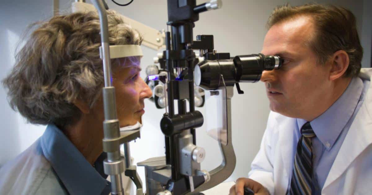
Fundoscopy (Ophthalmoscopic Examination)
A fundoscopic examination is performed to evaluate the retina, the optic disc, and the retinal vessels. It is done using an ophthalmoscope, a handheld illuminated lens device that allows the examiner to view a magnified image of the retina.
For a complete fundus exam, a mydriatic agent such as tropicamide may be used to relax the intraocular muscles and dilate the iris and pupil. Once the pupils are adequately dilated, the patient is placed in a darkened room and asked to focus on a fixed object on the wall behind the examiner. It is important to instruct the patient not to shift their gaze, as this would cause the intraocular structures to move and make the examination more difficult.
During fundoscopy, the ophthalmoscope is held in the examiner’s right hand, and the right eye is used to look through the viewing aperture when examining the patient’s right eye. When examining the left eye, the examiner uses the left hand and left eye. This positioning prevents direct face‑to‑face contact between examiner and patient and does require them to be in close proximity.
The examination is concluded by asking the patient to look directly at the light. This allows the examiner to assess the macula. Macular abnormalities include, for example, a “cherry‑red spot.”

For more information, it is recommended to visit the Eye Center at Al Batal Specialized Complex in Jeddah to obtain comprehensive guidance on visual acuity, color discrimination, peripheral vision testing, and fundoscopy.
See also: Is It Safe to Use Colored Prescription Contact Lenses for Vision Correction?


