

Med info
Glaucoma (blue water): causes, symptoms, and modern treatment methods at Batal Specialized Complex
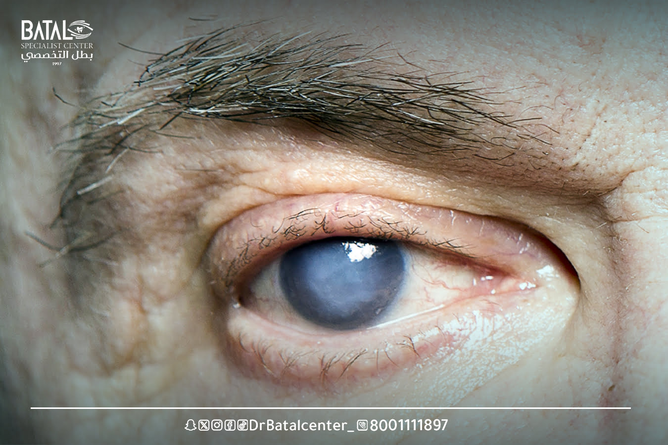
Glaucoma is the silent thief of sight. It affects the optic nerve in the eye and can lead to irreversible vision loss. It is believed that 4.5 million people worldwide are blinded by glaucoma, making it the third leading cause of blindness worldwide.
Eye conditions can be difficult to diagnose because symptoms don’t appear immediately but develop slowly over many years. This means that many patients only seek treatment when they notice they are losing their sight-that is, when the damage has already been done.
What is glaucoma (blue water)?
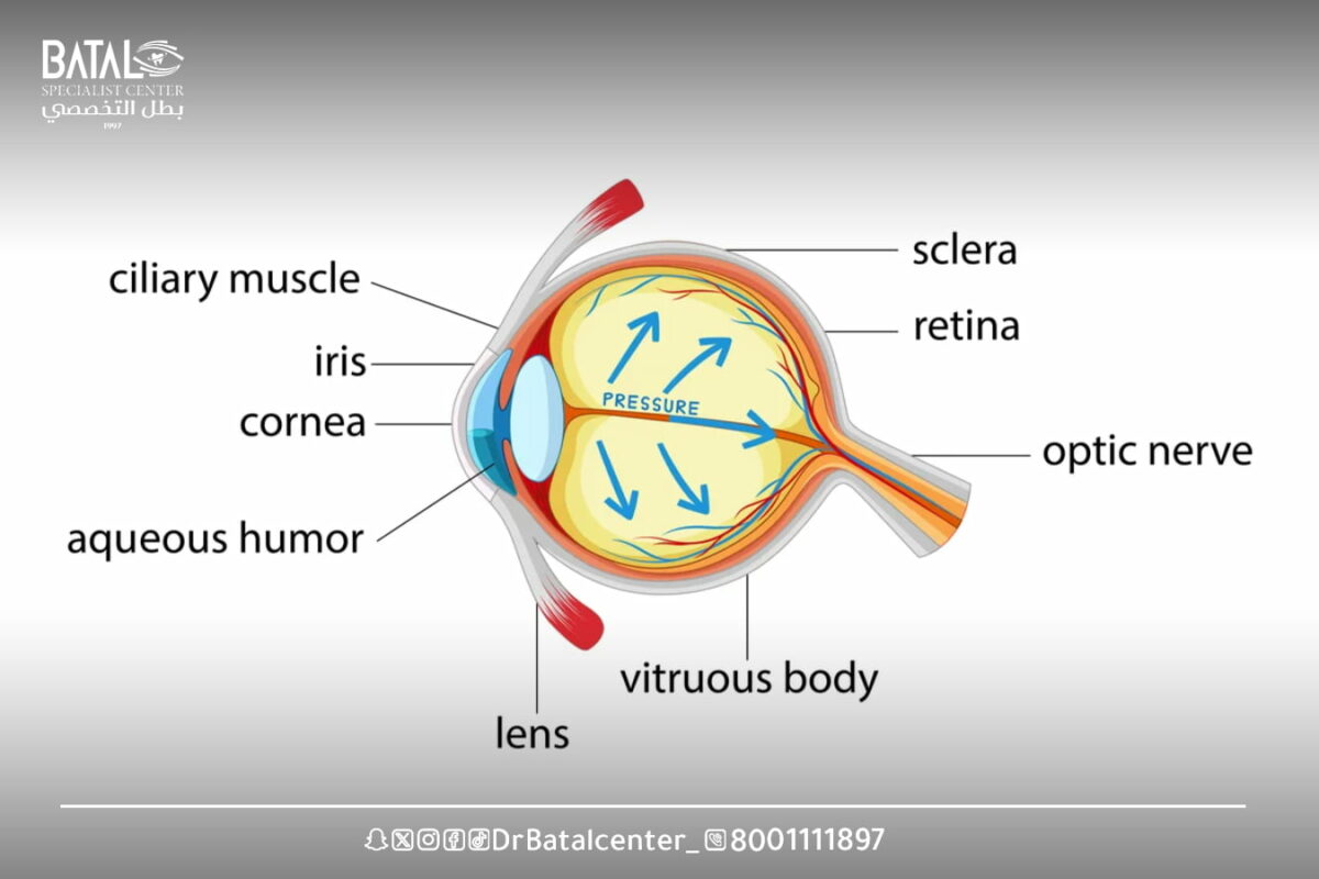
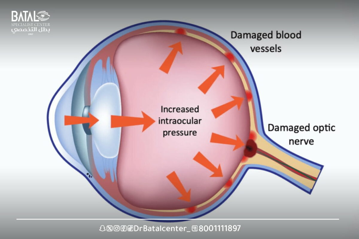
Glaucoma is an eye disease that gradually deteriorates vision by causing damage to the optic nerve that connects the eye to the brain as a result of the accumulation of pressure inside the eye.
Glaucoma usually has no symptoms in its early stages, and without proper treatment, glaucoma can lead to blindness. The good news is that with regular eye exams, early detection, and treatment, you can maintain your vision.
If you’ve been diagnosed with glaucoma, or suspect you have glaucoma, you probably have a lot of questions or concerns. Dealing with a long-term eye condition can seem stressful, but at Batal Specialist Medical Complex in Jeddah, we aim to help you learn how to maintain your eyesight and live a normal life with glaucoma.
Glaucoma in English
The medical name for glaucoma is Glaucoma
Types of glaucoma and the most important differences between them
Although there are many types of glaucoma, ophthalmologists usually classify it into two main categories: open-angle glaucoma and closed-angle glaucoma. Forms of glaucoma in both categories are characterized by damage to the optic nerve that can eventually lead to blindness.
Open-angle glaucoma
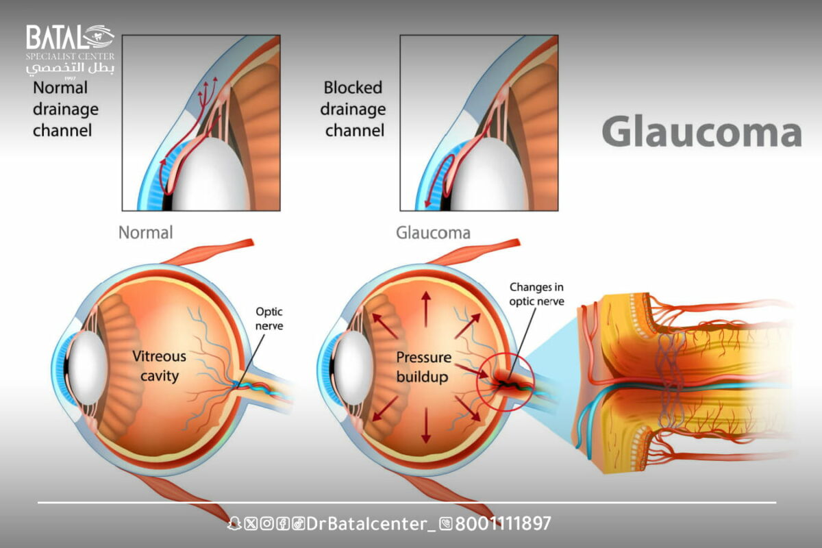
Primary open-angle glaucoma (POAG) is the most common form of glaucoma, in which internal intraocular pressure (or IOP) rises because the correct amount of fluid cannot be drained from the eye.
In primary open-angle glaucoma, the eye’s drainage angle is open but does not allow fluid to drain properly, and this clogs the eye’s drainage system over time, resulting in a slow increase in intraocular pressure.
This type of glaucoma is painless and often goes unnoticed until optic nerve damage becomes more serious, with some people losing their sight before they realize there is a problem.
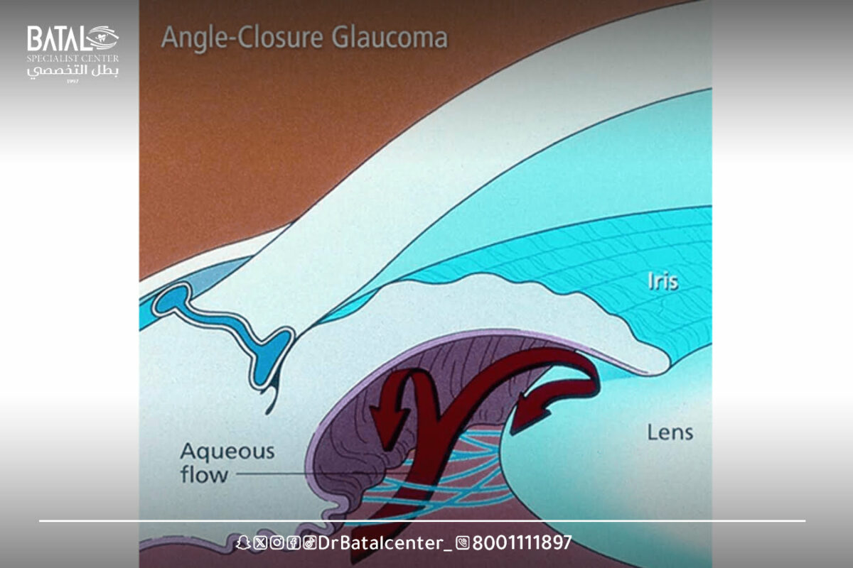
closed-angle glaucoma
Closed-angle glaucoma is a type of glaucoma in which the iris swells, and the swollen iris partially or completely blocks the drainage angle, and as a result, fluid cannot pass through the eye and pressure increases.
Closed-angle glaucoma is divided into acute closed-angle glaucoma or narrow-angle glaucoma, and it can occur suddenly or gradually.
Closed-angle glaucoma is rarer and very different from open-angle glaucoma, in which intraocular pressure usually rises very quick
Congenital glaucoma (in children)
Congenital glaucoma in children occurs when there is incorrect or incomplete development of the eye’s drainage channels during the prenatal period, and this is a rare and inherited condition, as glaucoma is diagnosed in children at the age of six months, and 80 percent of it is diagnosed in the first year of life.
Primary congenital glaucoma also develops from birth to 3 years of age, and juvenile open-angle glaucoma develops after 3 years of age.
Childhood glaucoma may also be caused by secondary causes such as eye injury and inflammation, or it may develop after cataract removal or after treatment with steroids.
Older children with pediatric glaucoma are usually asymptomatic like adults but may be able to describe visual discomfort such as sensitivity to light, loss of vision, problems adjusting to darkness, head or eye pain, and persistent eye redness.
Secondary glaucoma
Secondary glaucoma refers to any form of glaucoma in which there is a specific cause for increased intraocular pressure, leading to optic nerve damage and vision loss.
Secondary glaucoma can be of the open-angle or closed-angle type and can occur in one or both eyes.
Secondary glaucoma, also called neovascular glaucoma, may be caused by another eye disease or condition such as diabetes, asthma, chronic obstructive pulmonary disease (due to chronic steroid or corticosteroid use for more than three months), tumor, and cataracts. The type of treatment depends on the underlying cause, but usually includes medications, laser surgery, or conventional surgery.
Read more about: Tips for glaucoma patients
Causes of glaucoma and risk factors
The exact cause of glaucoma is not fully understood, but there are many factors that are believed to contribute to its development. Here are some of the main causes and risk factors associated with glaucoma:
High eye pressure
High eye pressure is one of the most important risk factors for glaucoma, as the fluid inside the eye is not drained properly, and it can accumulate, leading to increased pressure. This increased pressure can damage the optic nerve over time and cause vision loss.
Genetic factors
There is evidence that certain genetic factors play a role in the development of glaucoma. Studies have identified specific genes that may be associated with increased susceptibility to glaucoma. However, the inheritance pattern of glaucoma is often complex and can involve multiple genes.
Personal factors
Certain personal factors can increase your risk of developing glaucoma. These factors include age (people over 60 are at greater risk), race (people of African, Hispanic, and Asian descent are at greater risk for certain types of glaucoma), and family history of glaucoma.
Eye deformities
Structural abnormalities or anatomical differences within the eye can contribute to the development of glaucoma. These abnormalities can impede the normal flow of fluid and increase intraocular pressure. Examples include narrow-angle or closed-angle glaucoma, where the drainage angle between the iris and cornea is too narrow, or pseudoexfoliation syndrome, where abnormal protein deposits accumulate in the eye.
Secondary glaucoma
Secondary glaucoma refers to conditions in which glaucoma is caused by an underlying condition or injury. Various factors can contribute to secondary glaucoma, such as eye trauma, infections, certain medications (such as corticosteroids), eye tumors, diabetes, and some eye surgeries.
Early and advanced glaucoma symptoms
Signs and symptoms of glaucoma vary depending on its type and severity
How does a glaucoma patient see?
In open-angle glaucoma, there are no warning signs or obvious symptoms in the early stages, and most people don’t notice any change in their vision until the damage becomes very severe. However, when symptoms do occur, they include:
Intermittent blind spots around (lateral) or central vision that usually occur in both eyes.
- Blurred vision.
- Difficulty adapting to low light conditions.
- Symptoms of closed angle glaucoma include:
- Sudden onset of severe pain in the eye or forehead.
- Sudden loss of vision.
- Blurred vision.
- eye redness.
- Rainbow circles appear around bright lights.
- Headache.
- Nausea.
- Vomiting.
- Eye pain.
- Vomiting and nausea.
- halos around lights
- Glaucoma eventually leads to blindness if left untreated. At least 15% of people treated for glaucoma develop eye blindness.
Read more about: Eye redness: a comprehensive guide about it
How to diagnose glaucoma at Batal Specialist Complex
Diagnosis of glaucoma usually involves a comprehensive eye examination and various tests to evaluate optic nerve health, measure intraocular pressure (IOP), and evaluate peripheral vision. Here are some common diagnostic procedures and tests used to diagnose glaucoma:
Tension measurement: Tension measurement is a test used to measure eye pressure. This can be done using different methods, such as Goldman tonometry, where a small instrument gently touches the surface of the eye to determine pressure. High IOP is a major risk factor for glaucoma, but not all individuals with high IOP will develop this condition.
Ophthalmoscopy: Also known as fundus examination, it involves examining the optic nerve at the back of the eye. An ophthalmologist uses an ophthalmoscope to see the optic nerve, evaluate its appearance, and look for signs of damage or abnormalities associated with glaucoma.
Visual field testing: Glaucoma can cause loss of peripheral vision, so visual field evaluation is essential to diagnose and monitor the condition. A visual field test, such as automated perimeter measurement, measures a person’s ability to see objects in their peripheral (side) vision. It involves staring at a central point and responding when a light stimulus appears in different areas of the visual field.
Optical coherence tomography (OCT): A non-invasive imaging test that provides high-resolution cross-sectional images of the optic nerve and retina. It helps assess the thickness of nerve fibers and detect any structural changes or damage associated with glaucoma. OCT is particularly useful for monitoring disease progression over time.
Angleoscopy: Angleoscopy is performed to evaluate the angle of drainage of the eye. It involves using a special lens to examine the structures of the anterior chamber, including the angle at which the cornea meets the iris. It helps determine whether the angle is open or narrow, which is important in classifying types of glaucoma.
Corneal thickness measurement: Corneal thickness is crucial in accurately interpreting intraocular pressure measurements. Thin corneas may underestimate the actual IOP, while thick corneas may overestimate it.
Modern glaucoma treatment methods
The damage caused by glaucoma cannot be restored but the goal of treatment is to reduce intraocular pressure or pressure inside the eye. Treatment may include:
eye drops
Medications in the form of eye drops are often the first line of treatment for glaucoma. These eye drops work by either reducing fluid production inside the eye or increasing its drainage. Pharmaceutical eye drops that treat glaucoma include:
- Prostaglandin.
- Beta blockers.
- Alpha adrenergic agonists.
- Carbonic anhydrase inhibitors.
- Rho kinase inhibitor.
- Acuminate or cholinergic factors.
It is important to use eye drops as prescribed and regularly, as they help lower intraocular pressure and prevent further damage to the optic nerve.
Oral medications
In some cases, oral medications may be prescribed alongside or instead of eye drops to lower intraocular pressure. These medications work by reducing fluid production or improving fluid drainage. Oral carbonic anhydrase inhibitors are an example of such medications. Oral medications are usually used when eye drops alone are insufficient or not well tolerated. It may cause some side effects such as frequent urination, tingling in the fingers and toes, depression, upset stomach and kidney stones.
Laser treatment
Laser therapy can be used to treat glaucoma in different ways. One common procedure is called selective laser trabeculoplasty (SLT). It involves using a laser to enhance fluid drainage from the eye by treating the trabecular meshwork, the drainage system inside the eye.
Another laser procedure, called peripheral laser iridotomy (LPI), is used to treat narrow-angle or closed-angle glaucoma by creating a small hole in the iris to improve fluid flow. Laser treatment is usually performed in an ophthalmologist’s office and can be effective in lowering intraocular pressure (IOP).
Surgery
If medications and laser treatments are ineffective or inappropriate, various surgeries may be considered. Trabeculectomy involves creating a new drainage channel so that fluid can escape from inside the eye to lower pressure. Minimally invasive glaucoma surgery (MIGS) techniques are also available. Surgical interventions aim to improve fluid flow and reduce pressure inside the eye.
The importance of periodic follow-up with the ophthalmologist at Batal Complex
Regular follow-up with a doctor at Batal Complex is also crucial for individuals suffering from glaucoma. This allows the ophthalmologist to monitor the progression of the disease, evaluate the effectiveness of treatment, and make any necessary adjustments to the treatment plan.
Regular checkups also provide an opportunity to detect any changes in the visual field or optic nerve health, and adhering to a prescribed treatment regimen and attending follow-up appointments can help manage glaucoma effectively and reduce the risk of vision loss.
Glaucoma prevention and early detection
Although there is no guaranteed way to prevent glaucoma, there are some measures you can take to reduce the risk or delay its onset. In addition, early detection and prompt treatment are crucial in controlling the progression of glaucoma.
- Routine eye examinations are essential for early detection and diagnosis of glaucoma. A comprehensive eye exam is recommended at least once every two years, especially if you are over 40 or have other risk factors for glaucoma.
- Understanding your personal risk factors for glaucoma can help you be vigilant about your eye health.
- While lifestyle changes cannot guarantee glaucoma prevention, they can contribute to overall eye health. Maintain a healthy diet rich in fruits, vegetables, and omega-3 fatty acids.
- It is important to take steps to protect your eyes from injury, by wearing protective eyewear when engaging in activities that pose a risk of eye injury, such as sports or certain professions.
The importance of early detection and treatment in glaucoma control at the Batal Complex
The best way to prevent complications associated with glaucoma is through regular eye exams, early detection and immediate treatment at Batal Specialized Complex in Jeddah.
By detecting glaucoma in its early stages and managing it effectively, you can significantly reduce the risk of vision loss and maintain good eye health. It is important to visit the Batal Complex as they have the experience and equipment to perform comprehensive eye examinations and provide appropriate treatment options that suit your individual needs.
Learn about the blue water process at Batal Specialized Complex in Jeddah
At Batal Specialized Eye Complex, we have advanced experience in diagnosing and treating glaucoma (blue water) using the latest global devices and precise techniques in measuring eye pressure and examining the optic nerve.
Our medical team offers integrated treatment options including drug therapy, laser, and microsurgery for fluid drainage, with regular follow-up to ensure the condition is stable and vision is maintained safely.
Don’t wait for symptoms to appear, as glaucoma is a silent disease that can be controlled with early detection. Book a comprehensive eye examination now at Batal Specialized Complex in Jeddah and begin your journey to protect your eyesight with confidence.


