

Med info
Before and After Eye Treatment Photos
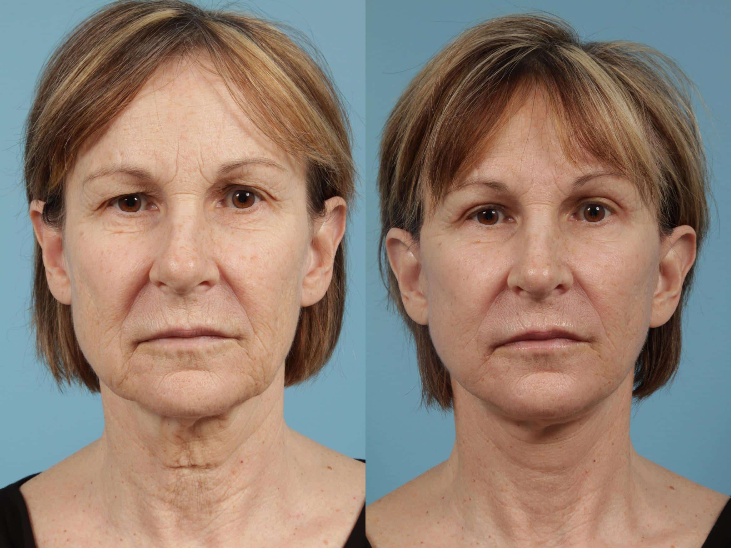
In this article, we will discuss common eye problems and share before-and-after eye photos after treatment, since eye conditions are among the most sensitive and complex health issues. They often start with improper handling of contact lenses and poor eye hygiene.
Amblyopia, also known as “lazy eye,” affects only about 2–3% of the population, but if left untreated, it can have a major impact on vision and quality of life.
Central vision usually fails to develop properly in one eye, which is what we call amblyopia.
There is a similar condition called strabismus (eye misalignment), which can sometimes lead to amblyopia. Another related condition is hemifacial weakness or paralysis of the facial nerve, known as the seventh cranial nerve.
This nerve controls facial expressions, eyelid movement, and the muscles of the forehead and neck.
There is also blepharitis, a common inflammation of the eyelids that is sometimes associated with bacterial eye infections, dry eye symptoms, or certain skin conditions such as rosacea.
Blepharitis has two main forms: anterior blepharitis, which affects the outer front part of the eyelid where the eyelashes attach.
There are many other well‑known eye diseases, some of them serious, but the positive news is that science and modern medicine have found effective treatments for most of these problems.
Before and After Strabismus Photos
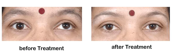
Before and After Strabismus Photos
Strabismus (eye misalignment) can be constant or intermittent. It usually occurs because the extraocular muscles that control eye and eyelid movement are not working together properly.
As a result, both eyes cannot focus on the same point at the same time. Strabismus can also result from an injury or a brain disorder that affects the eyes’ ability to work in coordination.
Persistent strabismus makes binocular vision impossible and causes loss of depth perception. It is estimated to affect about 2–5% of the general population.
Types of Strabismus
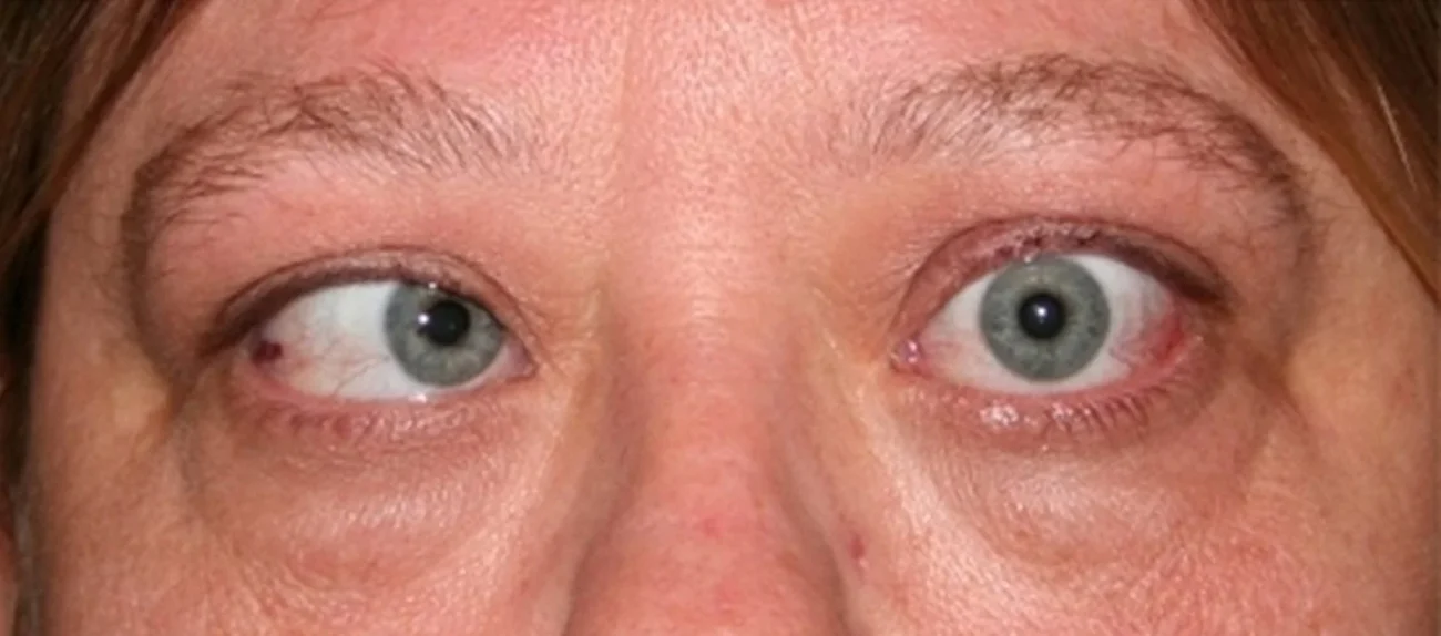
Types of Strabismus
There are different types of strabismus, which can be described according to their cause or the direction in which the eye turns. The following terms describe strabismus based on eye position:
– Hypertropia: this includes two situations—when the eye turns upward, and when the eye turns downward (often referred to more specifically as hypertropia for upward and hypotropia for downward deviation).
– Esotropia: when the eye turns inward.
– Exotropia: when the eye turns outward.
Early diagnosis of strabismus allows for more effective treatment. In the past, doctors believed that after a certain “critical period,” strabismus could no longer be treated.
While treatment is generally most effective up to around 6 years of age, strabismus surgery can actually be performed at any age.
Signs and Symptoms in Children
The signs of strabismus are usually fairly obvious from an early age, especially when one eye does not look straight ahead. Mild forms of strabismus can be less noticeable.
Infants and newborns may sometimes appear cross‑eyed, especially when they are tired. This does not necessarily mean they have true strabismus, but parents should still consult a doctor.
If one of the child’s eyes is often closed, or the child tilts their head when looking at objects, this may be a sign of double vision and possible strabismus. In such cases, it is best to see a doctor and contact us at Batal Specialized Center.
Strabismus is usually present at birth (congenital) or develops after about 3 months of age.
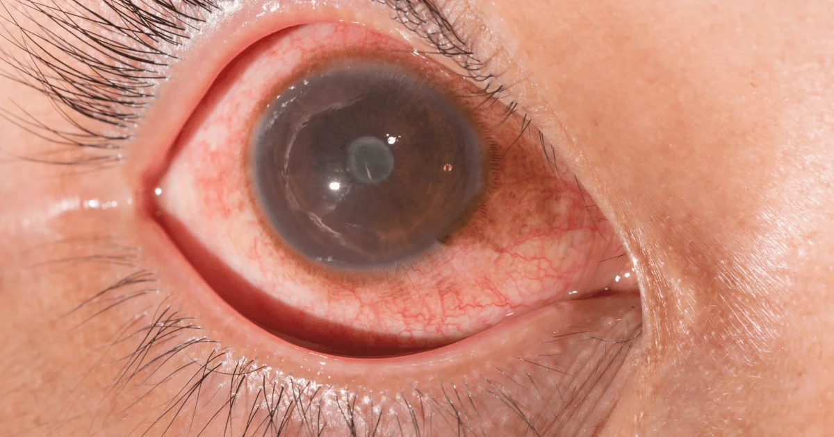
Before and After Glaucoma Photos
Before and After Glaucoma Photos
Your eye is constantly producing aqueous humor (the clear fluid inside the eye). As new fluid flows into the eye, an equal amount must drain out through a structure called the drainage angle.
This process keeps the pressure inside the eye (intraocular pressure) stable. If the drainage angle does not function properly, fluid builds up, eye pressure rises, and the optic nerve becomes damaged.
The optic nerve is made up of more than a million fine nerve fibers. It is similar to an electrical cable containing many tiny wires.
When these nerve fibers die, blind spots begin to appear in your field of vision. You might not notice these blind spots until most of the optic nerve fibers are already damaged. If all the fibers die, complete blindness occurs.
How Do You Develop Glaucoma?
How Does Glaucoma Develop?
There are two main types of glaucoma:
Open-Angle Glaucoma
This is the most common type of glaucoma. It develops gradually when the eye does not drain fluid as well as it should, much like a partially clogged drain.
As a result, eye pressure slowly increases and begins to damage the optic nerve. This type of glaucoma is painless and usually does not cause changes in vision at first.
Some people have optic nerves that are more sensitive to “normal” eye pressure, which puts them at a higher risk of developing glaucoma.
Regular comprehensive eye exams are essential to detect early signs of optic nerve damage.
Angle-Closure Glaucoma
This type occurs when the iris is very close to the eye’s drainage angle. The iris can end up blocking the drainage angle.
You can think of it like a sheet of paper sliding over a sink drain. When the drainage angle becomes completely blocked, eye pressure rises very quickly.
At that point, you must call your ophthalmologist immediately; otherwise, permanent vision loss or blindness can occur.
The signs of an acute angle-closure glaucoma attack include:
Many people with angle-closure glaucoma develop it slowly over time. This is called chronic angle-closure glaucoma. There are usually no early symptoms, so patients may not know they have it until significant damage has already occurred or they experience an acute attack.
Angle-closure glaucoma can lead to blindness if not treated right away.
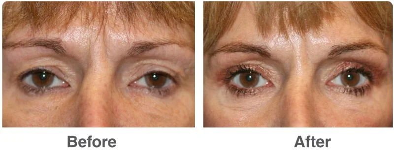
Read also: Upper Eyelid Lift Without Surgery – How It’s Done
Treatment of Glaucoma

Treatment of Glaucoma
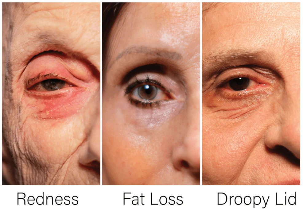
Glaucoma is usually controlled with prescription eye drops. These drops are used daily to lower eye pressure. Some work by decreasing the amount of aqueous fluid the eye produces.
Glaucoma medications can help preserve your vision, but they may also cause side effects. Some eye drops can cause:
Like all medications, glaucoma drugs may have side effects, and some may interact with other medications you are taking. That’s why it is important to give your doctor a complete list of all medicines you use regularly.
Be sure to talk with your ophthalmologist if you think you are experiencing side effects from your glaucoma medication. Never change the dose or stop using your glaucoma drops without consulting your eye doctor.
If you are about to run out of your medications, ask your ophthalmologist whether your prescription should be refilled.
Before and After LASIK Photos
Before and After LASIK Photos
LASIK (Laser-Assisted In Situ Keratomileusis) is a very common laser eye surgery, and there are many before-and-after eye photos that show the results. It can correct vision in people who are nearsighted, farsighted, or have astigmatism.
LASIK is one of several types of refractive surgery that work by reshaping the cornea—the clear front surface of the eye—so that light focuses properly on the retina at the back of the eye.
Read also: Eye Exam – Everything You Need to Know
Why Is LASIK Performed?
When light does not focus correctly on the retina, your vision becomes blurry. Doctors call this a “refractive error.” The main types of refractive errors are:
– Myopia (nearsightedness): you can see nearby objects clearly, but distant objects look blurry.
– Hyperopia (farsightedness): you can see distant objects more clearly, but nearby objects appear blurry.
– Astigmatism: this can make vision blurry at all distances because of an irregular shape of the eye or cornea.
Talk to your doctor about whether LASIK is suitable for you. You should not have this surgery if you:


