

Med info
Acanthamoeba Keratitis | Causes, Diagnosis, Treatment, and Complete Information 2026
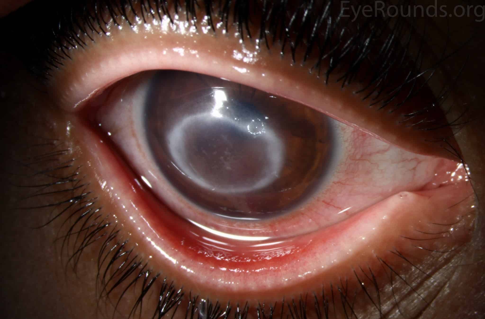
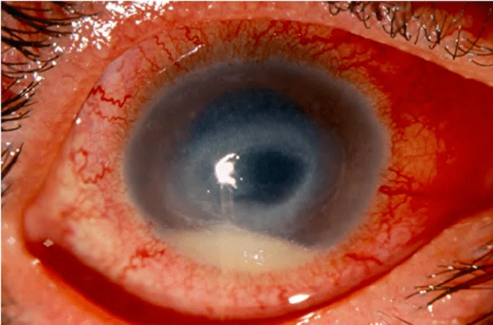
Acanthamoeba keratitis is a rare eye infection caused by the Acanthamoeba parasite. Doctors first identified it in 1974. The infection develops in the cornea, the clear front layer of the eye that covers the iris and pupil. Without prompt diagnosis and treatment, it can lead to permanent vision loss.
Contact lens wearers are at the highest risk of developing Acanthamoeba keratitis. Experts estimate that it affects about 1 to 33 contact lens users per million each year in developed countries.
Doctors often find Acanthamoeba keratitis difficult to diagnose because it can cause many of the same symptoms as other eye infections. Delayed diagnosis can result in more severe disease and complications.
What Causes Acanthamoeba Keratitis?
Keratitis is inflammation of the cornea, the transparent layer of tissue at the front of the eye that covers the pupil and iris. This particular form of keratitis develops when the cornea becomes infected by a single‑celled organism called Acanthamoeba.
Acanthamoeba is one of the most common free‑living organisms in the environment. It is found worldwide in soil and water. Most people are exposed to it, but only a small number develop disease. It can live in water sources such as:
– Tap water
– Swimming pools
– Hot tubs and Jacuzzis
– Lakes, rivers, and other freshwater sources
– Domestic water systems and air-conditioning units
The most common ways people develop Acanthamoeba keratitis are corneal trauma or exposure to contaminated water, such as using non‑sterile contact lens solution or submerging lenses in a hot tub or pool water.
Who Is at Higher Risk for Acanthamoeba Keratitis?
People who wear contact lenses are at the greatest risk of Acanthamoeba keratitis. Up to about 85% of those who develop the condition are contact lens wearers.
Acanthamoeba keratitis mainly affects middle‑aged adults. Individuals with weakened immune systems appear to be at increased risk, but the infection also occurs in people with normal immune function.
See also: Hyphema | Causes, Symptoms, and Treatment of Bleeding in the Eye
What Are the Symptoms of Acanthamoeba Keratitis?
Acanthamoeba keratitis usually affects one eye, but in rare cases, it can involve both eyes. Symptoms generally develop gradually over time.
The symptoms of Acanthamoeba keratitis are similar to those of many other eye infections and can include:
– Severe eye pain, often out of proportion to the clinical findings
– Redness of the eye
– Blurred or decreased vision
– Sensitivity to light (photophobia)
– Excessive tearing
– Foreign‑body sensation (feeling like something is in the eye)
– Eye irritation or burning
It can lead to complications such as:
– Corneal ulcers
– Corneal scarring and thinning
– Secondary glaucoma
– Permanent vision loss or blindness
How Do Doctors Diagnose Acanthamoeba Keratitis?
An ophthalmologist (eye specialist) can diagnose Acanthamoeba keratitis. However, diagnosis is often challenging because the disease is rare and cannot be identified based on symptoms alone.
Initially, physicians misdiagnose about 75% to 90% of cases. In a 2015 study, researchers found that nearly half of the people diagnosed with Acanthamoeba keratitis in Germany over a 10‑year period were first thought to have herpes simplex viral keratitis. Many studies do not clarify how much information patients shared with their doctors about contact lens hygiene and water exposure, even though ophthalmologists usually start the diagnostic process by examining your eye and taking a detailed medical and family history.
If they suspect Acanthamoeba keratitis, they may order:
– Corneal scraping for microscopic examination and culture
– Confocal microscopy (a special imaging test of the cornea)
– Corneal biopsy in selected cases
– PCR testing to detect Acanthamoeba DNA
See also: How Long Does It Take to Recover From Eyelid Surgery?
How Is Acanthamoeba Keratitis Treated?
Two medications, diamidines and biguanides, are commonly used as first‑line therapy. These are applied directly to the eye in the form of eye drops.
You’ll typically need to use the drops every hour during the first few days, then every 3 hours. Treatment often needs to be continued for 6 months to a year.
These two drug classes are effective for roughly 35% to 86% of patients. Most people receive them in combination to reduce the risk of drug resistance. The antibiotic neomycin may also be helpful when used together with other medications.
If medical therapy fails and the disease progresses to an advanced stage, you may need a corneal transplant (penetrating keratoplasty). In this surgical procedure, the surgeon replaces the damaged cornea with donor corneal tissue from a recently deceased person.
See also: Eyelid Lesions | Types, Symptoms, and Treatment
How Can I Reduce My Risk of Acanthamoeba Keratitis?

You can lower your risk of Acanthamoeba keratitis by avoiding contaminated water and preventing injuries to the cornea. Since most people who develop this infection wear contact lenses, if you use contact lenses you can reduce your risk by:
– Never rinsing or storing lenses in tap water
– Avoiding swimming, using hot tubs, or showering while wearing lenses
– Using only sterile, commercially prepared contact lens solutions

– Replacing lens cases regularly and letting them air‑dry
– Following your eye care professional’s instructions on lens wear and replacement schedules
– Avoiding overnight wear of contact lenses unless specifically prescribed


