

Med info
Everything You Need to Know About Eye Floaters | Causes and Treatment Options
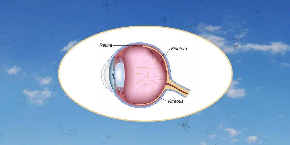
Eye floaters are spots or shadows that you may notice moving in your field of vision. They occur due to changes in the vitreous humor (the gel-like fluid) in the back of the eye, which can cast tiny shadows on the retina. In other words, eye floaters are deposits or clumps in the vitreous (often called the vitreous gel, vitreous humor, or vitreous body), the substance that fills the back part of the eye.
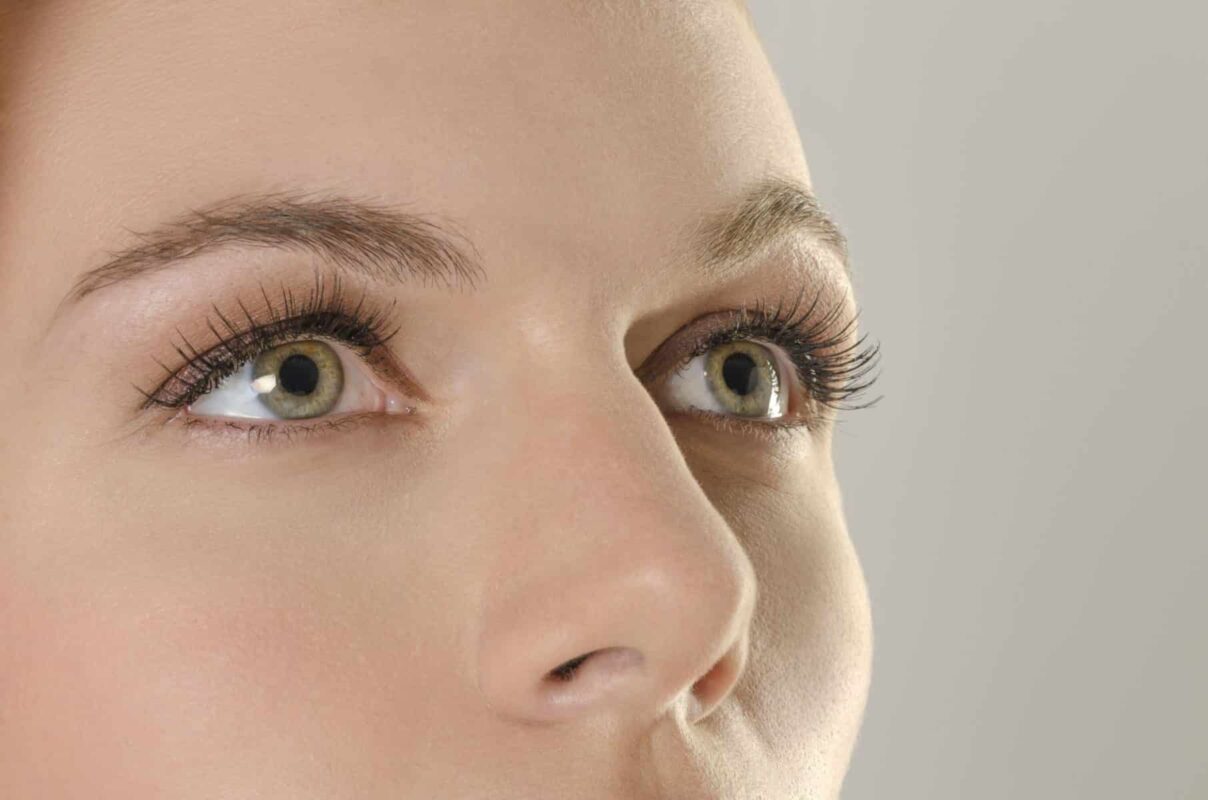
People use the term “eye floaters” because they see moving specks inside their vision that seem to drift when they move their eyes. Floaters may appear in just one eye or in both eyes.
How Do People Notice Eye Floaters?
The structures at the front of the eye (the cornea and the lens) focus light rays onto the retina, the light-sensitive tissue inside the eye. The light that reaches the retina must first pass through the vitreous humor, a gel-like substance that occupies about two-thirds of the back of the eye.
At birth and throughout childhood, the vitreous gel is usually clear and completely transparent. As we age, normal degenerative changes occur within the vitreous. Each of these changes in density creates tiny shadows on the surface of the retina. These shadows are what we perceive as eye floaters.
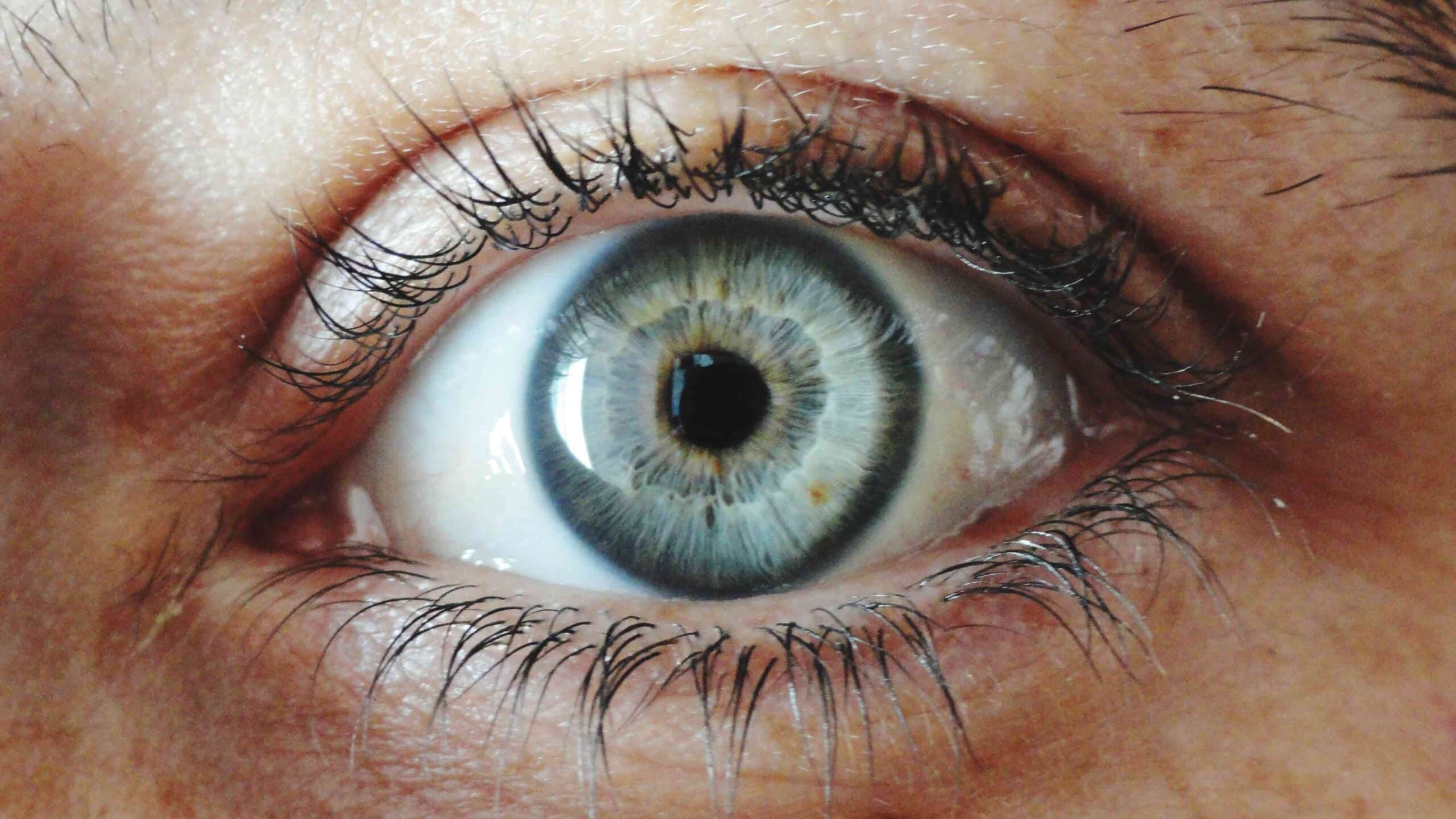
Floaters are usually light gray to black in color. As the eye moves from side to side or up and down, the shadows move as well and appear to drift or “swim” in the vision.

What Do Eye Floaters Look Like?
People describe eye floaters as black specks, small spots in their vision, straight or curved lines, cobwebs, strands, or ring-shaped or C-shaped spots. Some people see a single floater, while others may feel like they are seeing hundreds. The lines may be wavy, thick, or thin, and sometimes appear branched.
They can show up in varying shades of gray and are usually darker than the background you are looking at. The density and number of floaters vary from person to person and even within the same eye over time. Floaters are often more noticeable under certain lighting conditions, especially when looking at a bright, plain background, such as a clear sky or a white wall. They are rarely seen in dim lighting.
No two people have exactly the same floater pattern—much like fingerprints. If a person has floaters in both eyes, the pattern of floaters will be different in each eye and may change over time. Floaters always appear darker than the background and cannot be seen in total darkness or with the eyes closed. This is in contrast to flashes of light, which people often notice in the dark, even with their eyes closed.
What Causes Eye Floaters?
Most floaters are age-related. The changes usually begin within the vitreous gel itself. With aging, the vitreous naturally undergoes liquefaction (synchysis), forming small pockets of more liquid vitreous. In addition, the collagen fibers within the vitreous tend to thicken and clump together with age, creating floaters. Anyone over the age of 50 will have some degree of these changes, although the number and severity of floaters vary widely from person to person.
As the vitreous ages, the gel structure typically begins to shrink within the space it occupies. As it contracts, it can pull away from the retina and start to move more freely inside the eye, a process called posterior vitreous detachment (PVD). This can cause one or more large floaters, sometimes appearing as a large circular or ring-shaped shadow.
About 50% of adults around age 65 will have vitreous floaters in one or both eyes. If a PVD occurs in one eye, a similar event is more likely to happen in the other eye within the next 18 months. This is considered a normal part of aging.
However, many other abnormalities in the eye can also cause floater-like symptoms. Any cellular material suspended in the vitreous can create floaters, such as red blood cells from bleeding or white blood cells from inflammation.
Vitreous hemorrhage (bleeding into the vitreous cavity) can result from trauma, diabetic retinopathy, retinal tears involving blood vessels, or eye surgery. Inflammatory cells in the vitreous (vitritis) may be caused by infections, autoimmune eye diseases, penetrating or blunt eye injuries, or post-surgical inflammation.
Eye Diseases Associated with Abnormal Eye Floaters
Abnormal or sudden-onset floaters may be associated with:
– Diabetic retinopathy
– Retinal tears
– Retinal detachment
– High or pathological myopia (severe nearsightedness)
They are also more common in people who have had:
– Eye injuries
– Cataract surgery
– Laser eye procedures (such as certain retinal laser treatments)
Several inflammatory or infectious conditions are linked to floaters, including:
– Tuberculosis-related eye disease
– Sarcoidosis
– Syphilis
– Toxoplasmosis
– Acute retinal necrosis
A rare condition called asteroid hyalosis (calcium-lipid deposits in the vitreous) is another cause of floaters.
Primary or secondary intraocular tumors, including ocular lymphoma and leukemia, can be associated with floaters as well, although these causes are extremely rare.
Risk Factors for Developing Eye Floaters
Major risk factors for developing eye floaters and related complications include:
– Increasing age (the main risk factor)
– Myopia (nearsightedness), especially high myopia, as posterior vitreous detachment tends to occur at a younger age in highly myopic individuals
– Diabetes mellitus, due to the risk of diabetic retinopathy and vitreous hemorrhage
Loss of peripheral (side) vision can be a warning sign of retinal detachment, which may be associated with new floaters and/or flashes of light.
How Are Eye Floaters Diagnosed?
If new floaters suddenly appear, or if existing floaters suddenly increase in number, an immediate evaluation by an ophthalmologist is essential. An ophthalmologist is a medical doctor specializing in eye care and can provide comprehensive assessment and treatment. This includes:
– Performing a complete eye examination
– Prescribing eyeglasses or contact lenses if needed
– Diagnosing and treating eye diseases
– Performing eye surgery
– Evaluating visual function and related problems
It is especially important that the ophthalmologist examines the entire eye, including the vitreous and the retina.
When a patient presents with floater symptoms, the ophthalmologist will begin by taking a detailed medical and ocular history. They will then:
– Test central and peripheral vision
– Examine the front of the eyes using a slit-lamp microscope
– Instill dilating eye drops to widen the pupils
– Examine the vitreous and retina using bright light and special lenses, often with a head-mounted indirect ophthalmoscope
Based on this examination, the ophthalmologist can determine whether there are any abnormalities—such as retinal tears, retinal detachment, or inflammation—that require further testing or urgent treatment.
Are Eye Floaters Treatable?
Most common age-related floaters tend to become less bothersome over time. This happens for several reasons:
– The floater material may be partially broken down or absorbed through natural processes within the eye
– Floaters may shift to a position where they cast less noticeable shadows on the retina
– The brain gradually adapts to their presence and “tunes them out,” making them less noticeable in day-to-day life
For many people, floaters decrease in apparent size and intensity, or become easier to ignore, even if they do not disappear completely.
Can Medication Help Get Rid of Eye Floaters?
Many herbal remedies, nutritional supplements, home treatments, and iodine-containing products have been promoted as natural cures for eye floaters. However, none of these have been proven effective in well-designed clinical trials.
Anti-inflammatory medications or antibiotics may reduce the number of white blood cells in cases where floaters are caused by inflammation or infection, but:
– There is no proven oral medication
– There is no eye drop
that can reliably reduce or eliminate the common, age-related type of eye floaters.
Can Surgery Remove Eye Floaters?
Some ophthalmologists have proposed treating floaters with a YAG laser to break them up. However, there is currently no strong evidence that this procedure is both safe and consistently effective. Using a laser in this way also carries significant potential risks to vision.
Another option is surgical removal of the vitreous gel itself, in a procedure called a vitrectomy. During a vitrectomy, the vitreous is removed and replaced with a clear solution. While vitrectomy can reduce or eliminate floaters, performing it solely for common age-related floaters due to posterior vitreous detachment involves substantial risks, including:
– Cataract formation or rapid progression of existing cataracts
– Retinal detachment
– Intraocular bleeding
– Infection inside the eye (endophthalmitis)
Because of these risks, vitrectomy is usually reserved for severe, vision-threatening situations or when floaters are extremely disabling and other causes have been ruled out.
Is It Possible to Prevent Eye Floaters?
There is no known way to completely prevent age-related eye floaters or to make them disappear on demand. However, certain measures can help reduce the risk of floaters caused by other conditions:
– Wearing safety glasses or protective eyewear when using power tools or engaging in activities that risk eye injury
– Tight blood sugar control in people with diabetes to reduce the risk of diabetic retinopathy and vitreous hemorrhage

In general, learning from others’ experiences—what many describe as “my experience with eye floaters”—can provide practical tips on coping strategies, when to seek urgent care, and how to adapt to floaters in daily life. Nevertheless, any sudden change in floaters, appearance of light flashes, or loss of peripheral vision requires prompt evaluation by an ophthalmologist.


