

Med info
Tear Ducts: Anatomy, Function, and Common Conditions
The tear ducts, also called the nasolacrimal ducts, are channels that run along both sides of the nose and connect the eyes to the nasal cavity, creating a pathway through which tears can drain. While this system may not seem very complex, the tear ducts play a vital role in maintaining the health of your eyes. That’s why infections, blockages, and other problems affecting them can be a serious concern for your eye health.
The process begins in the lacrimal glands, which are located in the upper outer corner of the eye socket on each side. When you blink, your eyelids spread the tears across the surface of the eye. From there, tears drain into the puncta—tiny openings located at the inner corners of the upper and lower eyelids. Tears then pass through small drainage channels called canaliculi, which lead into the lacrimal sac attached to the side of the nose. The lacrimal sac sits in a bony groove called the lacrimal fossa and empties into the nose through the nasolacrimal duct. At that point, the tears either evaporate or are reabsorbed.
See also: Conjunctivitis: Types, Causes, and Treatment
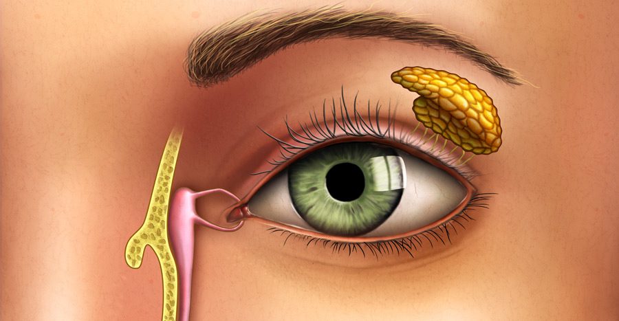
Anatomy of the Tear Duct
The nasolacrimal duct lies alongside the nose and represents the final step in the eye’s drainage system. Each time you blink, the eyelids distribute tears produced by the lacrimal glands over the surface of the eye. The tears are then collected into tiny drainage openings called puncta. There are both upper and lower puncta, and both are found at the inner corner of each eye.
From the puncta, tears flow into narrow channels called canaliculi (pronounced “can-uh-LICK-you-lye”). These canaliculi drain into the lacrimal sac located next to the upper part of the nose. The lacrimal sac then tapers down into the nasolacrimal duct, which carries the tears through the bone of the nose and into the nasal cavity.
See also: Causes of Blurred Vision and Related Conditions
Function of the Tear Ducts
The nasolacrimal duct works by draining “used” tears from the surface of the eye into the nose. From there, the tears are either absorbed, wiped away with a tissue, or pass into the throat and are swallowed. The tear-producing glands in your eyes make different types of tears for different purposes. Some types of tears keep the eye lubricated, while others help wash away irritants and foreign particles.
On average, a person produces about 15 to 30 gallons of tears every year, all of which need to be drained through the tear ducts. When a tear duct becomes blocked, tears cannot drain properly. This can lead to pain, swelling, inflammation, and infection around the inner corners of the eyes.
See also: Yellowing of the Eyes: Causes, Treatment, and When to Seek Care
Conditions Affecting the Tear Duct

Duct Obstruction
Tear duct obstruction (nasolacrimal duct obstruction) can also be present at birth, a condition known as congenital nasolacrimal duct obstruction. Because tears cannot drain normally, blocked tear ducts can cause tears to back up, leading to pain along the tear duct and epiphora (excessive tearing). Tears that cannot drain as they should may overflow down the face, or become trapped and stagnant inside the duct. This can result in a swollen, inflamed tear duct, which can be painful.
Dacryocystitis (Lacrimal Sac Infection)
If left untreated, bacteria, fungi, or viruses can grow within stagnant tears and cause an infection of the lacrimal sac, a condition known as dacryocystitis. Dacryocystitis may be acute or chronic. Acute dacryocystitis develops rapidly, with pronounced and often severe symptoms, but it typically responds well to treatment with antibiotic ointments or oral antibiotics, and symptoms usually improve shortly after starting therapy.
Chronic dacryocystitis causes milder symptoms that develop gradually but may persist for months. Topical steroid eye drops are often prescribed in chronic cases to help reduce inflammation and swelling.
In some people with chronic dacryocystitis, surgery on the tear duct system may be recommended. This procedure, called dacryocystorhinostomy (DCR), creates a new drainage pathway. The surgeon uses instruments, often including a laser, to clear blockages and remove small pieces of bone around the tear duct. The result is a wider channel that allows tears to drain properly.
Protecting the Tear Ducts

Al Batal Specialty Center is equipped with highly advanced technologies for diagnosing and treating eye problems and for checking visual acuity and eye health. These include procedures such as Presbyond and ReLEx SMILE (femtosecond SMILE) for treating different degrees of refractive errors, as well as MicroPulse laser technology for managing eye misalignment and certain types of strabismus. You can book an appointment at Al Batal Eye Center for a comprehensive eye examination performed by some of the most experienced ophthalmologists in Jeddah.


