

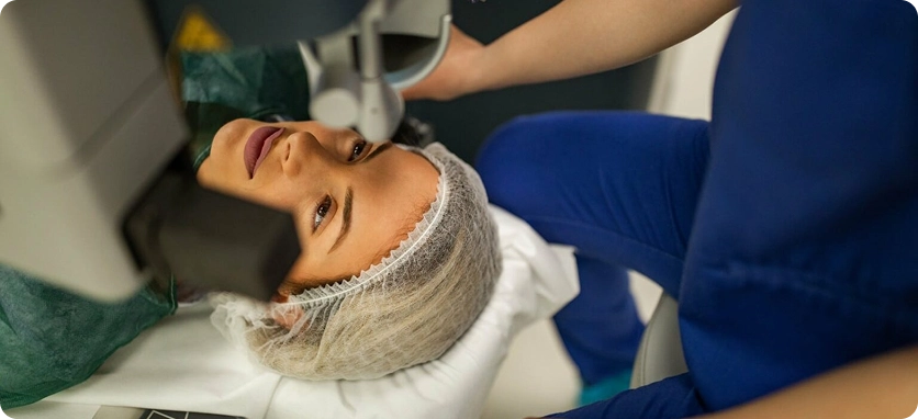
Ocular Ultrasound Imaging in Jeddah
Ocular ultrasound imaging is one of the most advanced diagnostic tools used at Batal Specialist Medical Centerr to evaluate eye conditions with high accuracy. This non-invasive and completely safe test can be performed for adults, children, and infants, providing fast and precise diagnostic insights without any surgical intervention. We continuously invest in advanced imaging technologies to support accurate diagnosis, personalized treatment plans, and comprehensive eye care according to the highest medical standards.

Ocular Ultrasound Imaging in Jeddah
Ocular ultrasound imaging is one of the most advanced diagnostic tools used at Batal Specialist Medical Center to evaluate eye conditions with high accuracy. This non-invasive and completely safe test can be performed for adults, children, and infants, providing fast and precise diagnostic insights without any surgical intervention. We continuously invest in advanced imaging technologies to support accurate diagnosis, personalized treatment plans, and comprehensive eye care according to the highest medical standards.

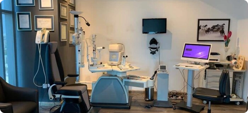
Wide-Field Ocular Ultrasound System
Batal Specialist Medical Center is equipped with a state-of-the-art ocular ultrasound system capable of imaging both the anterior and posterior segments of the eye, providing detailed evaluation of the retina and vitreous body. The system also features advanced international calculation formulas for precise measurement of intraocular lens (IOL) power for cataract surgery, ensuring highly accurate lens selection and optimal visual outcomes.
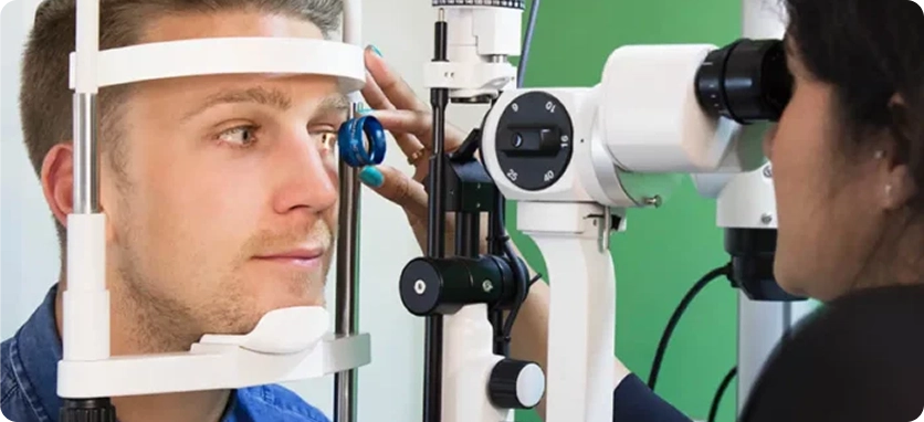
What Is Ultrasound Imaging?
Ultrasound imaging is a painless, non-invasive diagnostic method that uses high-frequency sound waves to examine the body and the eye. When sound waves pass through tissues and organs, they reflect in different ways, allowing specialists to create detailed internal images for accurate diagnosis. Unlike X-rays, ultrasound does not use radiation and is completely safe.
In most cases, no special preparation is required before the ultrasound exam. The patient lies comfortably on an examination table, and a clear water-based gel is applied to the eye area to help transmit the sound waves. A handheld probe (transducer) is then gently moved over the area being examined.
There is no radiation risk, and patients can resume their normal daily activities immediately after the test.
When Is Ocular Ultrasound Used at Batal Specialist Medical Center
Ocular ultrasound is a simple, painless procedure that typically takes between 5 and 20 minutes. The eye does not need to remain open during the test, and no special preparation is required beforehand. Children usually do not need anesthesia, making it a safe and convenient diagnostic option for all age groups.
For these reasons, we use ocular ultrasound imaging in many clinical situations to support accurate diagnosis and effective treatment planning.
Ultrasound for Eye Examination
We use ocular ultrasound imaging when direct examination with a slit lamp and fundus examination is not sufficient. This advanced diagnostic tool helps in the rapid detection of serious eye conditions that may require urgent intervention, such as retinal detachment, vitreous hemorrhage, and lens dislocation.
Who Is a Candidate for Eyelid Surgery?
If aging has caused changes in the size or shape of your eyes, you may be a good candidate for eyelid surgery.
Eyelid cosmetic surgery (blepharoplasty) is suitable for individuals who are bothered by sagging eyelid skin, under-eye bags, wrinkles, and fat deposits that make them look older than their actual age.
Patients considering eyelid surgery should be in good overall health and free from serious eye conditions to ensure safe and optimal results.
Sound Waves for Surgical Monitoring
Ultrasound imaging is also one of our essential tools for monitoring treatment progress and surgical outcomes. It uses harmless sound waves with no known side effects and is cost-effective, allowing repeated use during treatment to accurately assess the patient’s response and recovery.
Identifying the Type of Eye Bleeding
Eye bleeding may appear as red spots inside the eye, redness of the white part of the eye, or even blood-tinged tears in some cases. Ocular ultrasound examination helps determine the type and cause of eye bleeding, allowing specialists to plan the appropriate treatment.
Diagnosing Eye Conditions
In routine eye examinations, light-based tests are usually sufficient. However, traditional eye exams can be limited in cases such as intraocular bleeding or dense cataracts. In these situations, ocular ultrasound imaging plays a crucial role in detecting and diagnosing eye conditions before determining the appropriate treatment plan.
This advanced imaging test is used to detect and evaluate:
Eye tumors or cysts
Measurement of tumor size and thickness in suspected cancer cases
Retinal detachment
Glaucoma diagnosis and evaluation
Cataracts
Post-LASIK and post-IOL implantation follow-up (after cataract surgery)
Foreign bodies inside the eye, such as blood clots, masses, or lipid cysts on the iris
Eye swelling or bulging (proptosis)
Orbital injuries (eye socket trauma)
Vitreous detachment
Axial length measurement of the eye
Benefits of Ocular Ultrasound Imaging
Ultrasound imaging offers several advantages that make it a preferred diagnostic tool in eye care:
High-frequency sound waves help evaluate soft tissues that are not visible on X-ray imaging.
No exposure to ionizing radiation, making it safer than other diagnostic techniques such as X-rays and CT scans.
More cost-effective compared to many other imaging methods.
Painless procedure with no injections, needles, or anesthesia required in most cases.
The examination takes only a few minutes.
Immediate results for faster diagnosis and treatment decisions.
The test can be performed with the eyelids closed, making it ideal for children and patients with light sensitivity or discomfort.
In most cases, anesthetic drops are not required. However, if the examination needs to be performed with the eyes open, the doctor will apply numbing eye drops to ensure patient comfort before starting the procedure.
Ultrasound Imaging | Doppler System
Ultrasound technology has advanced significantly and now includes specialized Doppler imaging, which uses unique sound waves to measure blood flow through blood vessels in a fast and painless way. Doppler ultrasound helps detect vascular problems and blood clots that may not be visible with standard ultrasound imaging.
Doppler ultrasound is a relatively advanced technique and has proven to be a promising tool for evaluating vascular eye diseases. It provides valuable diagnostic insights for conditions such as optic nerve disorders, diabetic retinopathy, and glaucoma.
Doppler ultrasound testing provides our specialists with critical information to determine the most effective treatment approach. You can ask your doctor whether Doppler ultrasound affects eye color—generally, it does not.
Types of Doppler Ultrasound Examinations
Color Doppler: Measures the speed and direction of blood flow in real time.
Power Doppler: An advanced form of Color Doppler that provides more detailed blood flow information but does not show flow direction; Spectral Doppler is often used for directional analysis.
Spectral Doppler: Helps evaluate the degree of blood vessel obstruction and is similar to Duplex Doppler.
Duplex Doppler: Measures blood flow in blood vessels and provides structural and flow information.
Continuous Wave Doppler: Allows highly accurate measurement of high-velocity blood flow.
Conditions Diagnosed with Doppler Ultrasound
Doppler imaging helps diagnose:
Eye blood clots
Eye swelling (ocular edema or bulging)
Weak blood vessels and fluid accumulation behind the eye
How to Book an Ocular Ultrasound Examination
You can book an ultrasound examination at the reception desk at Batal Eye Specialty Center, and you won’t have to wait long before being called by our specialized technician.
Alternatively, you can schedule your appointment online at a time that suits you.
Ocular and Orbital Ultrasound Results
Your ophthalmologist will review the ultrasound results with you and ensure that the eye measurements obtained from the scan are within the normal range.
B-scan ultrasound provides your doctor with detailed structural information about your eye. If the results are abnormal, your doctor will investigate the underlying cause. Conditions that may be detected with a B-scan include:
Foreign bodies inside the eye
Cysts
Swelling or edema
Retinal detachment
Tissue damage or orbital (eye socket) injury
Vitreous hemorrhage (bleeding into the gel-like substance that fills the back of the eye)
Retinal cancer, subretinal tumors, or other eye tumors
What Is Ocular Ultrasound?
Ocular ultrasound uses high-frequency sound waves to measure and produce detailed images of the eye and the orbit (the bony cavity in the skull that holds the eye).
This test provides more detailed visualization of the internal structures of the eye compared to a routine eye examination. It is usually performed by a trained ultrasound technician or an ophthalmologist (a specialist in diagnosing and treating eye diseases).
Ocular ultrasound studies can be performed in a clinic, imaging center, or hospital setting, depending on the case and available facilities.
Why Do I Need an Ocular Ultrasound?
Your ophthalmologist may recommend ocular ultrasound if you have unexplained eye problems or if you have recently experienced trauma or injury to the eye area.
This procedure is useful for identifying eye abnormalities and diagnosing various eye conditions. Some of the issues that ocular ultrasound can help detect include:
Eye tumors or masses
Foreign bodies in the eye
Retinal detachment
Ocular and orbital ultrasound can also be used to diagnose or monitor conditions such as:
Glaucoma (a progressive disease that can lead to vision loss)
Cataracts (clouding of the natural lens)
Intraocular lens implants (IOLs) — artificial lenses implanted after removal of the natural lens, usually due to cataracts
Do You Need an A-Scan or B-Scan Ultrasound?
A-Scan Ultrasound
A-scan ultrasound uses high-frequency sound waves to measure the length of the eye, corneal curvature, and to calculate the appropriate intraocular lens power before cataract surgery. The test can be performed while sitting or lying down.
B-Scan Ultrasound
B-scan ultrasound helps visualize and examine the structures behind the eye. In some cases, cataracts or other eye conditions make it difficult to see the back of the eye using routine examination tools. In such cases, ultrasound imaging plays a key role in diagnosing tumors, retinal detachment, and other eye conditions.
Our Doctors – Batal Specialist Medical Center
Dr. Ahmad Hassan Batal
Senior Consultant, Pediatric Surgery and Eye Surgery Fellow of the British Royal Surgical College Fellow of the American Academy of Ophthalmology
Dr. Wadah Chalabi
Member of the British Royal College of Surgeons<br>Vitreoretinal and cataract surgery
Dr. Baraa Faham
Dr. Issa Baissa
Consultant in Ophthalmology & Vision Correction
Dr. Mohamad Jeed
15 years of experience in pediatric eye diseases and strabismus.
Dr. Obaida Kilani
Eye Surgeon specializing in Glaucoma (Water Glaucoma) Surgery, Cataract Surgery, and Advanced Laser Vision Correction Procedures.
Dr. Aziz Al-Balawi
Saudi Board of Ophthalmology<br>
Diseases and surgery of the Retina, Vitreous and laser
Dr. Tarek Al Najjar
more than 25 years of experience in eyes Oculoplastic surgeries and the lacrimal system.
Dr. Mohamed hantira
the certificate of the Scientific Council of Ophthalmology Glaucoma, laser vision correction and corneal surgery
Dr. Mohammed Al-Mousa
Consultant in Corneal Surgery, Vision Correction & Cataract
Get comprehensive medical care


Installments available up to 100% via Tamara & Tabby & Amwal


